Overview
A condition that is characterized by pain in the front part of the knee and surrounding the patella (kneecap) is called patellofemoral pain syndrome (PFPS). Doctors also refer to this condition as “jumper’s knee” or “runner’s knee.”
The condition is not serious, despite leading to symptoms ranging from sore to very painful kneecap. Just rest and conservative treatment measures can also lead to pain reduction in patients affected by this disorder.
Causes of Patellofemoral Pain Syndrome
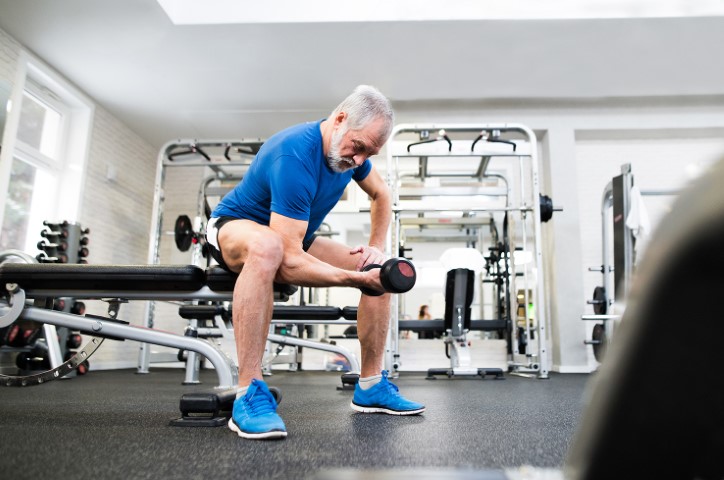
- Knee Overuse: In many cases, PFPS is caused by vigorous physical activities that put repeated stress on the knee —such as jogging, squatting, and climbing stairs. It can also be caused by a sudden change in physical activity. This change can be in the frequency of activity—such as increasing the number of days you exercise each week. It can also be in the duration or intensity of activity—such as running longer distances.


Other factors that may contribute to patellofemoral pain include:
- Use of wrong sports training techniques or equipment
- Altered sizing in footwear or playing surface
- Wrong Alignment of the Kneecap: Patellofemoral pain syndrome can also be triggered by irregular tracking of the kneecap in the trochlear groove. In this condition, the patella is protruded to one side of the groove upon knee bending. This deviation may lead to elevated pressure between the rear of the patella and the trochlea, irritating the soft tissues.
Factors that add to poor kneecap tracking include:
- Malalignment of the legs between the hips and the ankles. Malalignment may lead to shifting of kneecap which is very far to the outside or inside of the leg, or it may ride very high in the groove of the trochlea—leading to patella Alta.
- Muscular disparities or weaknesses, particularly in the front thigh muscles (quadriceps). During knee bending and straightening, the quadriceps muscles and tendon aid in keeping the kneecap inside the trochlear groove. Weak or imbalanced front thigh muscles can lead to poor kneecap tracking inside the groove.
Any individual can be affected by this condition, however, athletes more frequently encounter this problem.
Symptoms of Patellofemoral Pain Syndrome
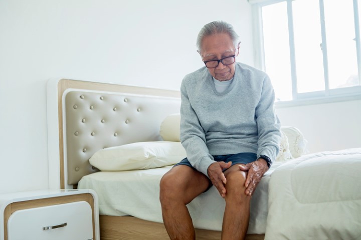
The sure sign symptom of patellofemoral syndrome is a dull, aching pain usually occurring on the front portion of the knee or around the kneecap. The pain may be felt in one or both knees and often deteriorates with movement.


Other painful symptoms are:
- Pain during exercise
- Pain during knee bending, including navigation through stairs, jumping or sitting on your heels
- Pain after sitting for a prolonged period with bent knees
- Cracking or popping sounds in the knee during stair navigation or post sitting for a prolonged duration
Diagnosis of Patellofemoral Pain Syndrome
Your doctor will inquire about your knee history and apply pressure on your knee areas. He/she will also move your leg in various positions to help exclude other conditions, having similar signs and symptoms.
To determine the exact cause of your knee pain, your doctor may prescribe imaging tests like:
- X-rays: A small quantity of radiation enters your body to create X-ray images of your body on a screen. Though this tool sees bone well, it is less effective at enabling soft tissue visualization.
- CT scans. This technique combines images of X-ray from different angles to form thorough images of internal structures. Though CT scans can envisage both bone as well as soft tissues, the procedure carries a much higher radiation dose as compared to plain X-rays.
- MRI: MRIs create thorough images of bones and soft tissues, such as the knee ligaments and cartilage using a combination of radio waves and a strong magnetic field. However, they are much more costly as compared to the above diagnostic tools.
Treatment of Patellofemoral Pain Syndrome
In many patients, simple home remedies can improve pain in patients affected by this condition.
1) Treatments at home
Changes in daily routine
Discontinue performing activities that hurt your knee, till your pain is sorted out, including alterations in your exercise routine or moving to low-impact exercises. Latter activities may apply less stress on your knee joint. If your body weight is above normal, dropping weight also helps in reducing pressure on your knee.
RICE Method
Opting for the RICE method may go a long way in improving your state. RICE is an acronym for Rest, Ice, Compression and Elevation.
- Rest: Avoid applying weight on the knee which is paining.
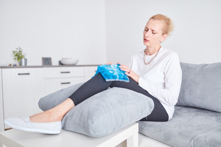
- Ice: Apply ice packs for 20 minutes at a single time, multiple times in a single day.


- Compression: Cover the knee lightly with an elastic bandage, leaving a cavity in the kneecap area, to avoid extra swelling. Ensure that the bandage is loose enough to not cause any pain.
- Elevation: Rest with your knee elevated to a height above your heart, as frequently as possible.
Medicines
Taking certain NSAIDs like ibuprofen or naproxen can aid in the reduction of swelling and pain. If your pain continues or worsens, preventing knee motion, kindly contact your doctor. Medical treatment for patellofemoral pain syndrome is meant to relieve pain and re-establish the range of knee motion and its strength. In the majority of cases, this pain can be treated by non-surgical means.


2) Non-surgical Treatment
Physical Therapy
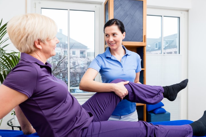
Dedicated exercises for improving range of motion, strength, and knee joint endurance can contribute to providing relief in this condition. It is particularly vital to concentrate on strengthening and stretching your front knee muscles as these muscles are the key stabilizers of your kneecap. Experts may also recommend core muscle exercises to build up the muscles in your abdomen and lower back.


Using Orthotics
Orthotics/shoe inserts can contribute to alignment and stabilization of your foot and ankles, withdrawing stress from your lower leg. These devices can either be customized or bought directly.
3) Surgical Treatment
Surgery is very rarely required to treat patellofemoral pain syndrome, only in extreme cases, which are not responding to non-surgical modes of treatment.
Arthroscopy
During this procedure, your surgeon inserts a small camera, referred to as an arthroscope, into your knee joint. The camera shows images of the knee joint on a screen, which your surgeon uses to guide small surgical instruments in the area to be operated on.
- Debridement: In some cases, removal of damaged smooth, white tissue covering the ends of bones at the joints (articular cartilage) from the kneecap’s surface can give relief from pain.
- Lateral release: If the affected muscle is very tight to pull the kneecap out of the trochlear groove, this procedure can relax the tissue and resolve the improper alignment of the kneecap.
- Tibial tubercle transfer: In some cases, kneecap realignment by shifting the patellar tendon along with a portion of the bony prominence on the shinbone may be essential.
A conventional open surgical cut is needed in this procedure. The surgeon detaches the tibial tubercle, partially or completely, to enable shifting the bone and the tendon to the knee’s inner side. The piece of bone is then reattached to the shinbone using screws. In the majority of cases, this transfer permits better kneecap tracking in the groove of the trochlea.
Realignment
In more extreme cases, a surgeon may require operating your knee for re-alignment of your kneecap’s angle or to reduce pressure on the cartilage.
To conclude, Patellofemoral pain syndrome is one of the most common causes of anterior knee pain today. And, while there are several treatment options available to treat the syndrome, it is crucial to accept the fact that your knees are absorbing a huge amount of pressure since the time you have started walking. Plus, with regular wear and tear, knee pain is bound to happen and take a toll over a while as even the knee’s two shock absorbers — pads of cartilage called menisci — start to deteriorate with age. But certain steps like regular stretching and mobility drills, wearing good footwear, and practicing correct form while exercising should be paramount if you are looking to age-proof your knees.


