Category

Cancer

Category

What if we could catch a major women’s health threat early enough to truly make a difference? Today, we are championing a cause that could change the course of many lives by empowering every woman to take control of her health through regular ovarian cancer screening. As we explore this critical topic, let’s uncover how accessible and essential health practices can help detect this silent killer early.

What is Ovarian Cancer?

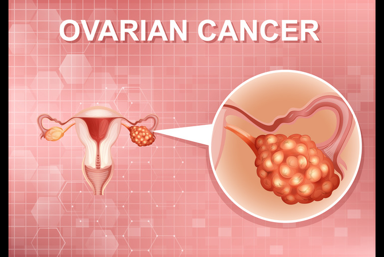
Ovarian cancer begins in the ovaries, which is where the eggs are produced. This cancer often goes unnoticed in its early stages because it typically doesn’t show many clear or specific symptoms, which can make it hard to detect early. When symptoms like bloating, pelvic pain, and changes in bathroom habits do occur, they often resemble common stomach and digestive issues, leading many to overlook them until the cancer has progressed. As per the World Cancer Research Fund, globally, ovarian cancer is the eighth most common cancer in women.

Types of Ovarian Cancer Screening

Screening for ovarian cancer typically involves methods designed to detect cancer before symptoms become apparent. The most common screenings include the transvaginal ultrasound (TVUS) and the CA-125 blood test. TVUS is a type of pelvic ultrasound used extensively to examine the reproductive organs and detect anomalies in the ovaries. Meanwhile, the CA-125 blood test measures the level of the protein CA-125, which is often elevated in women with ovarian cancer.

Who is Eligible for Ovarian Cancer Screening?

The following groups of women are recommended to undergo regular screenings based on their risk levels:

Women with Genetic Predispositions: Those who have a family history of ovarian, breast, or colorectal cancer should consider starting screening earlier and more frequently. This group may benefit from annual screenings starting from the age of 30 or 10 years earlier than the youngest age at the diagnosis in the family, whichever comes first.

Women with a Personal History of Cancer: Those who have previously had breast, uterine, colon, or rectal cancer often have a higher risk of developing ovarian cancer and should discuss their screening schedule with their healthcare provider.

Postmenopausal Women: Older women, especially those who have gone through menopause, are at a higher risk for ovarian cancer. They should discuss the feasibility and timing of screenings like TVUS and CA-125 blood tests with their healthcare professionals.

Regular Interval Discussions: For those who are at increased risk, discussing the appropriate intervals for screening with their healthcare provider is crucial. Generally, annual screenings are recommended, but the frequency may increase based on individual risk assessments and the advice of their healthcare provider.

Overcoming Barriers to Screening

Despite the availability of screening methods, various barriers deter women from undergoing regular ovarian cancer screenings. Lack of awareness about the disease and its screening options tops the list, followed by psychological barriers such as fear of diagnosis. Cultural beliefs and societal norms also play a significant role, where discussing gynecological health may be taboo or stigmatized. Moreover, in regions where healthcare is not readily accessible or affordable, even willing individuals may find it challenging to seek help.

Empowering women to overcome these hurdles involves a multifaceted approach. Raising awareness through community education, creating supportive environments that respect cultural sensitivities, and enhancing access to healthcare services are vital steps. Additionally, encouraging open conversations about gynecological health and normalizing these discussions can significantly reduce stigma and promote a more proactive attitude towards health screening.

Importance of Advocacy and Education

The role of advocacy and education in enhancing awareness about ovarian cancer cannot be overstated. By informing women about the importance of early screening and its potential to save lives, we can shift the narrative from fear and ignorance to empowerment and action. Advocates and educators can influence public health policies, improve healthcare services, and ensure that every woman understands the risks and the benefits of early detection.

Wrapping It Up

Let us renew our commitment to ensuring no woman is left behind in the fight against ovarian cancer. Through increased understanding, accessible screening, overcoming societal barriers, and strong advocacy, we can make a significant difference. Every woman must be empowered to take charge of her health by making ovarian cancer screening a priority.

https://www.wcrf.org/cancer-trends/ovarian-cancer-statistics/
https://www.cdc.gov/cancer/ovarian/basic_info/screening.htm
https://www.mayoclinic.org/tests-procedures/ca-125-test/about/pac-20393295#:~:text=Overview,high%20risk%20of%20the%20disease.
https://ocrahope.org/get-the-facts/statistics/#:~:text=Ovarian%20cancer%20accounts%20for%202.5,the%20deadliest%20of%20gynecologic%20cancers.
https://pubmed.ncbi.nlm.nih.gov/33620837/#:~:text=Ovarian%20cancer%20is%20the%20leading,poor%20outcomes%20of%20this%20disease.
https://www.ncbi.nlm.nih.gov/pmc/articles/PMC6459072/
https://www.cancer.org/cancer/types/ovarian-cancer/detection-diagnosis-staging/detection.html
https://www.cancerresearchuk.org/about-cancer/ovarian-cancer/getting-diagnosed/screening
https://www.uptodate.com/contents/screening-for-ovarian-cancer-beyond-the-basics#:~:text=In%20this%20group%2C%20ovarian%20cancer,125%20and%20a%20pelvic%20ultrasound.

Laparoscopy

Laparoscopy is a minimally invasive surgery in which an incision is made near the belly button. A thin tube with a fitted camera, also known as a laparoscope, is inserted. The camera allows the surgeon to see the organs in the pelvic area and locate the endometriosis. The surgeon can then remove the endometriosis tissue using a small surgical instrument.

Laparoscopy is usually recommended for mild to moderate endometriosis. It is a safe and effective procedure with a faster recovery time than open surgery. Patients can get back to their routine within a week.

The benefits of laparoscopy for endometriosis surgery are:

  • Minimally invasive: Laparoscopy requires only tiny incisions, resulting in less scarring and a faster recovery time.
  • Accurate: The camera allows the surgeon to see the endometriosis tissue and remove it precisely.
  • Cost-effective: Laparoscopy is less expensive than open surgery.
  • Shorter hospital stay: Laparoscopy usually is an outpatient procedure; the patient can go home very next day of the procedure.

Hysterectomy

A hysterectomy is a surgical intervention that involves the removal of the uterus. In certain instances, the procedure may also include the removal of the fallopian tubes and ovaries. Hysterectomy is usually recommended for severe endometriosis that does not respond to other treatments.

Hysterectomy can be done differently; vaginal, abdominal, and laparoscopic. The type of surgery depends on the patient’s condition and the surgeon’s preference. 

The benefits of hysterectomy for endometriosis surgery are:

  • Permanent solution: Hysterectomy removes the uterus, which means the patient will no longer have periods and endometriosis-related pain.
  • Effective: Hysterectomy is a highly effective treatment for severe endometriosis.
  • Reduced risk of ovarian cancer: Removing the ovaries reduces the risk of ovarian cancer in women with a high risk of developing it.
  • Improved quality of life: Hysterectomy can improve the patient’s quality of life by relieving pain and other symptoms.

Excision Surgery

In excision surgery, the endometriosis tissue is removed from the affected organs. This surgery is usually recommended for women with severe endometriosis.

Excision surgery can be laparoscopic or an open surgery, depending on the extent of the endometriosis. The surgeon removes the tissue using a surgical instrument and then repairs any organ damage.

The benefits of excision surgery for endometriosis surgery are:

  • Precise removal of endometriosis tissue: Excision surgery removes the endometriosis tissue completely, reducing the recurrence risk.
  • Preserves fertility: Excision surgery can preserve the ovaries and fallopian tubes, improving the patient’s chances of conceiving.
  • Improved quality of life: Excision surgery can relieve pain and other symptoms, improving the patient’s quality of life.

Radical Hysterectomy

Radical excision surgery is recommended for women with severe endometriosis that has spread to other organs in the pelvic area. It involves removing the affected organs, such as the uterus, ovaries, fallopian tubes, and sometimes even the bladder or rectum.

Radical excision surgery can be done as an open surgery or laparoscopically, depending on the extent of the endometriosis. The surgeon removes the affected organs and then repairs any damage to surrounding tissues.

The benefits of radical excision surgery for endometriosis surgery are:

  • Permanent solution: Radical excision surgery removes the affected organs, which means the patient will no longer have periods and endometriosis-related pain.
  • High success rate: Radical excision surgery has a high success rate in treating severe endometriosis.
  • Reduced risk of recurrence: Removing the affected organs reduces the risk of recurrence.
  • Improved quality of life: The patient’s quality of life may be improved by undergoing radical excision surgery, which can alleviate their pain.

Conclusion

In conclusion, endometriosis surgery can help manage the symptoms of endometriosis and improve the patient’s quality of life. However, the recommended type of surgery depends on the endometriosis’s severity and extent. Each type of surgery has its benefits. Patients must consult their doctor to discuss the various options and determine the best treatment plan for their individual needs.

References

https://www.healthline.com/health/endometriosis/laparoscopy-for-endometriosis
https://my.clevelandclinic.org/health/treatments/4620-endometriosis-surgery
https://www.brighamandwomens.org/obgyn/infertility-reproductive-surgery/endometriosis/surgical-treatment-of-endometriosis-excision-and-destruction#:~:text=Excision%20of%20endometriotic%20lesions%20or,different%20kinds%20of%20energy%20sources.
https://www.circlehealthgroup.co.uk/health-matters/womens-health/radical-endometriosis-surgery#:~:text=What%20is%20radical%20surgery%20for,other%20treatments%20did%20not%20help.

What Is Rectal Cancer?
Rectal cancer develops in the rectum, a chamber between the end of the large intestine and the anus. It usually starts as a growth on the inner lining of the rectum and can spread to other body parts if left untreated. Symptoms of rectal cancer may include changes in bowel habits, rectal bleeding or blood in the stool, abdominal discomfort, and unexplained weight loss.

Diagnosis and Staging of Rectal Cancer
Diagnosing rectal cancer involves a combination of tests and procedures that help identify cancer cells in the rectum. Standard diagnostic tests include a digital rectal exam, colonoscopy, CT scan, MRI, and biopsy. These tests help determine the location and size of cancer and whether it has spread to nearby tissues or organs. In addition, the staging of rectal cancer is crucial in determining the most appropriate treatment options. The TNM staging system is used to stage rectal cancer based on tumour size, lymph node involvement, and distant metastases.

Treatment Modalities for Rectal Cancer in India
The treatment of rectal cancer typically involves a combination of surgery, radiation therapy, and chemotherapy. The choice of treatment modality depends on the cancer stage, the tumour’s location, and the patient’s overall health. The treatment modality includes- 

  • Surgery: Surgery is the most common treatment modality for rectal cancer. It is a highly effective treatment, mainly when the cancer is detected early. The benefits of surgery include the removal of cancerous tissue and the prevention of cancer recurrence. It involves the removal of the cancerous tumour along with surrounding tissues and lymph nodes. The type of surgical procedure used depends on the stage and location of cancer.
  • Radiation Therapy: Radiation therapy is a treatment modality that uses high-energy radiation to kill cancer cells. It is usually applied with surgery to kill any remaining cancer cells that may not have been removed during surgery. The benefits of radiation therapy include the destruction of cancer cells and the prevention of cancer recurrence.
  • Chemotherapy: As a treatment modality, chemotherapy uses drugs to kill cancer cells. It is typically used with surgery and radiation therapy for advanced-stage rectal cancer or when cancer has spread to other body parts. Chemotherapy drugs can be administered orally or intravenously. It can help shrink tumours, prevent cancer recurrence, and slow the progression of advanced-stage cancer. Chemotherapy can also improve the patient’s quality of life by relieving symptoms and reducing pain.

Choosing the Right Treatment Modality for Rectal Cancer
Choosing the suitable treatment modality for rectal cancer depends on several factors, including the cancer stage, the tumour’s location, the patient’s overall health, and preferences.

Conclusion
Rectal cancer is a significant health concern in India, and early detection and treatment are crucial for successful outcomes. With early detection and proper treatment, rectal cancer can be successfully treated, and patients can go on to live healthy and fulfilling lives.

FAQs
Q: What is the most common treatment modality for rectal cancer?
A:
 Surgery is the most common treatment modality for rectal cancer. Different surgical procedures may be used depending on the stage and location of the tumour.

Q: Can chemotherapy be a standalone treatment modality for rectal cancer?
A:
 Chemotherapy is rarely used as a standalone treatment modality for rectal cancer. It is usually used with other treatments, such as surgery and radiation therapy, to improve outcomes.

Q: Which factors should be considered when selecting a treatment modality for rectal cancer?
A:
 Several factors should be considered when selecting a treatment modality for rectal cancer, including the stage of cancer, the location of the tumour, the patient’s overall health, and the patient’s preferences and values.

Reference links:
https://www.cancercenter.com/cancer-types/colorectal-cancer/types/care-at-city-of-hope#:~:text=Rectal%20cancer%20occurs%20when%20cells,with%20colorectal%20cancer%20is%2068.
https://www.cancer.gov/types/colorectal/screening-fact-sheet
https://www.cancer.org/cancer/colon-rectal-cancer/treating/rectal-surgery.html

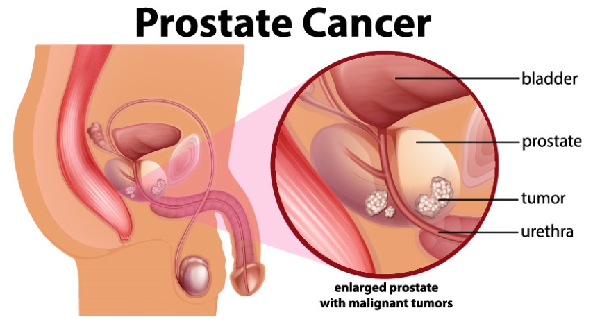
The prostate gland is a small gland found only in males. It produces the seminal fluid that helps transport the sperms. The prostate gland is located below the bladder and in front of the rectum. As the name itself suggests, prostate cancer begins at the prostate gland, when the cells grow and multiply uncontrollably.

Prostate cancer is one of the most common types of cancer, and it may show no signs or symptoms in the early stages. However, patients with prostate cancer have higher chances of complete recovery if the cancer is detected early.

Symptoms of Prostate Cancer

As mentioned above, prostate cancer does not show any signs or symptoms during the earlier stages. Some common symptoms visible during the advanced stage of prostate cancer are:

  • Trouble urinating, or the need to urinate more often than usual
  • Blood present in the urine
  • Blood in the semen
  • Erectile dysfunction
  • Pain in the bones – especially the hips, spine, and ribs
  • A feeling of weakness/numbness in the feet
  • Loss of weight

While most of these signs and symptoms may point to several other underlying conditions or diseases, it is necessary to talk to your healthcare provider if these symptoms are persistent or continue to worsen.

What causes prostate cancer?

Though the causes of prostate cancer remain unclear, it is known prostate cancer occurs when the cells in the prostate mutate. The mutation causes cells to grow and multiply rapidly, forming abnormal cells. The abnormal cells continue living, while the healthy cells die. These growing abnormal cells form a tumor and invade the nearby tissues or organs. Gradually, these abnormal cells also spread to the other parts of the body, causing cancer to grow.

Risk Factors of Prostate Cancer

Several factors can increase the risk of prostate cancer, such as:

Age: The risk of prostate cancer increases with age. Prostate cancer is more prevalent among men over the age of 50.

Ethnicity: For reasons unknown and undetermined, prostate cancer is more common among men of African-American descent. As per to urology.org, African-American men tend to get diagnosed with prostate cancer at an earlier age, and the cancer is likely to be aggressive in growth.

Family History: Men with a family history of prostate cancer have higher chances of being diagnosed with the same. A strong family history of breast cancer can also increase the chances of one developing prostate cancer.

Obesity: People who are obese have higher chances of developing prostate cancer, compared to those who maintain a healthy weight.

Prostate Cancer Diagnosis

If your doctor suspects that you might have prostate cancer, he might enquire about your symptoms such as urinary and sexual problems. He might also ask you about possible risk factors such as your family history to analyze the chances of you developing prostate cancer. after this, the doctor will run several diagnostic tests to diagnose the disease:

Digital Rectal Exam (DRE): During this, the doctor might insert a gloved finger into your rectum to examine the prostate, and feel for any bumps or hardened areas. If the doctor finds any abnormalities in the shape or size of the gland, he may advise further tests.

PSA Blood Test: A blood sample is taken and tested for PSA. PSA stands for Prostate-specific Antigen, which is a protein produced by the prostate gland. It is normal for a small amount of PSA to be found in your bloodstream. However, if it is higher than usual, it may be an indication of prostate cancer.

Ultrasound: During this test, a small probe is inserted into the rectum, and uses soundwaves to create images of the prostate gland.

MRI: Magnetic Resonance Imaging (MRI) scan may be suggested by your doctor to create a more detailed picture of your prostate gland.

Biopsy: The doctor may also collect a sample of cells in your prostate gland to check for signs of cancerous cells. This procedure is known as a prostate biopsy. A thin needle is inserted into the prostate to collect a sample of the tissue.

If you are diagnosed with prostate cancer, the doctor may use tests such as ultrasound, CT scan, or MRI to determine the stage of cancer.

Treating Prostate Cancer

The treatment for prostate cancer depends on how aggressive the cancer is and the extent it has spread. The right treatment option will also be chosen depending on the side effects and their impact on overall health.

Surgery: This involves removing the prostate gland, along with some nearby tissues and lymph nodes. Surgery is usually performed when the cancer is confined to the prostate. It may also be used to treat advanced prostate cancer combined with other treatment methods.

Radiation Therapy: This involves using high-powered radiation to kill the cancerous cells. This can be performed externally (external beam radiation) or internally (brachytherapy), by placing radiation sources in the prostate tissue.

Cryotherapy: This treatment involves using extremely cold gases to freeze the prostate tissue. After this, the tissue is allowed to thaw, and the procedure is repeated.

Hormone Therapy: This treatment stops the body from producing testosterone, the male hormone. Since prostate cancer cells rely on testosterone to grow, cutting the hormone supply may cause the cancerous cells to die.

Chemotherapy: This therapy uses drugs to kill the rapidly growing cancerous cells. Chemotherapy is usually used when cancer has spread to the other organs and areas of the body.

Immunotherapy: As the name itself suggests, this therapy involves using the immune system to fight cancer cells. The immune system usually does not attack cancer cells as they produce proteins that help protect the cancer cells. Immunotherapy interferes with this process.

Targeted Therapy: This treatment emphasizes the abnormalities present in the cancerous cells. Targeted therapy blocks and combats these abnormalities, causing the cancer cells to die.

Though several factors increase the risk of developing prostate cancer, early detection and following a healthy lifestyle can help nip prostate cancer in the bud. Though certain risks such as age, family history, and ethnicity cannot be avoided, other factors such as obesity can be avoided by maintaining a healthy diet and exercising regularly. Talk to your healthcare provider immediately if you notice certain symptoms and take the necessary precautions!

Lung cancer is the most common cancer in the world. According to the World Health Organization, there were 2.21 million cases in the year 2020. It is the most common cause of death around the world. Patients have a 13 times better chance of living for five years if they are diagnosed early. Lung Cancer Awareness Month (LCAM) is celebrated every year in November to raise awareness about the disease and continue to challenge the stigma associated with lung cancer.

The goal of the month is to urge individuals to seek medical assistance sooner rather than later, to encourage early diagnosis so that patients have the best chance of a successful treatment, and to emphasise other key aspects that affect patient outcomes. Educating people on the complexity of lung cancer, the vast spectrum of people who are affected, and the harmful effects of lung cancer stigma can aid in earlier diagnosis and better patient treatment. Furthermore, LCAM is also an occasion to highlight therapeutic developments, advocate for global access to care, and, most importantly, demonstrate our support for patients and their loved ones.

Facing the stigma

Lung cancer is often misunderstood to be solely a smoker’s disease. However, more than half of individuals diagnosed are former smokers or non-smokers. This misunderstanding has been associated with poor outcomes due to factors like waiting too long to seek treatment, disease-related distress, a lack of social support, and poor care quality.

What you need to know about Lung cancer

Cancer is a condition in which the cells of the body grow out of control. Lung cancer is cancer that starts in the lungs and spreads throughout the body. Lung cancer begins in the lungs and can spread to the lymph nodes or other organs in the body, including the brain. Cancer that has spread to other organs may extend to the lungs as well. Metastases are the spread of cancer cells from one organ to another.

There are two types of lung cancer:

Small cell lung cancers (SCLC) and non-small cell lung cancers (NSCLC) are the two forms of lung cancer. This classification is based on the appearance of tumour cells under a microscope. Making the distinction between these two types of tumours is critical since they develop, spread, and are treated differently.

SCLC accounts for roughly 10% to 15% of all lung malignancies. This form of lung cancer is the most aggressive and fastest-growing of all. Cigarette smoking is highly linked to SCLC. SCLCs spread quickly throughout the body, and they are usually detected after they have spread widely.

The most common type of lung cancer is non-small cell lung cancer (NSCLC), which accounts for roughly 85% of all occurrences. NSCLC is divided into three categories based on the cells detected in the tumour. They are as follows:

  • Adenocarcinomas, like other lung cancers, are linked to smoking, this form is also seen in nonsmokers, particularly women, who get lung cancer. The majority of adenocarcinomas develop in the lungs’ periphery. They have a proclivity for spreading to lymph nodes and beyond.
  • Squamous cell carcinomas used to be more common than adenocarcinomas, but now they make up around 25% to 30% of all lung cancer cases. Squamous cell tumours are most common in the bronchi of the central chest. This type of lung cancer tends to stay in the lung, spread to lymph nodes, and grow large enough to produce a cavity.
  • Large cell carcinomas, also known as undifferentiated carcinomas, are the least prevalent kind of NSCLC, accounting for 10% to 15% of all lung cancer cases. This malignancy has a significant proclivity for spreading to lymph nodes and distant locations.

Types of Treatment

Depending on the type of lung cancer and how far it has gone, there are numerous treatment options. Surgery, chemotherapy, radiation therapy, targeted therapy, or a combination of these treatments can be used to treat non-small cell lung cancer. Small cell lung cancer is usually treated with a combination of radiation and chemotherapy.

  • Surgery: A procedure in which surgeons remove cancerous tissue from the body.
  • Chemotherapy: Special medications are used to shrink or kill cancer cells. The drugs can be pills that you consume or medicines that are injected into your veins, or both.
  • Radiation therapy: Treatment that involves the use of to kill cancer, high-energy rays (similar to X-rays) are used.
  • Target therapy: Drugs are used to stop cancer cells from growing and spreading. The drugs can be taken orally or administered intravenously. Before targeted therapy is employed, tests will be performed to determine if it is appropriate for your cancer type.

In the treatment of lung cancer, doctors from various specialties frequently collaborate. Pulmonologists are doctors who specialise in lung illnesses. Surgeons are medical professionals who carry out procedures. Thoracic surgeons specialise in surgery of the chest, heart, and lungs. Medical oncologists are doctors who specialise in using medications to treat cancer. Radiation oncologists are doctors who use radiation to treat cancer.

It is important to educate yourself and your loved ones about the most common type of cancer so that lives can be saved. If you notice anything unusual contact your doctor immediately.