Category

Surgery:

Category

Introduction

Coronary artery disease (CAD) is a serious condition that affects millions of people around the world. It is a form of cardiovascular disease that is caused by the hardening and narrowing of the arteries that supply blood to the heart. If left untreated, ‍coronary artery disease (CAD) lead to chest pain, complete blockage of an artery, heart attack, and cause sudden death. Knowing the symptoms, risk factors, and treatments for CAD can help you protect yourself and those around you from its potentially devastating effects. In this article, we’ll explore what you need to know about coronary artery disease and how to identify and manage this devastating condition and keep it at bay. However, there are also various preventative measures that you can take to reduce your risk of developing the condition. With the right information and knowledge, you can reduce your risk of developing this dangerous condition and improve your overall heart health.

What is coronary artery disease?

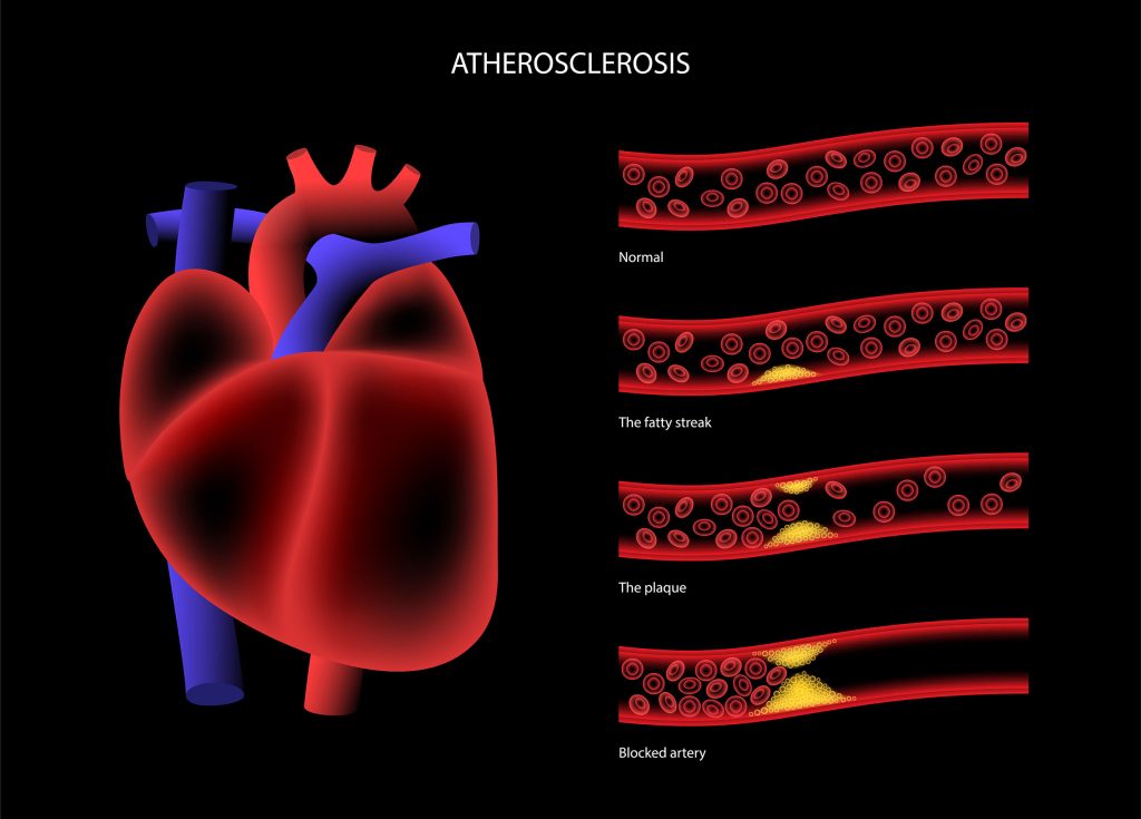
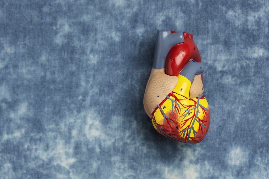
Coronary artery disease (CAD) is a medical condition in which plaque builds up in the arteries that supply blood to your heart.  The buildup of fatty deposits in the arteries is a common precursor to CAD, this build-up of fatty deposits in the arteries could be formed over a period of time, which can completely block the arteries and cause heart failure. This buildup causes the arteries to narrow, which increases your risk of a heart attack. The plaque that builds up in the arteries can be made of cholesterol, calcium, and pieces of fatty material. It usually starts in the walls of the arteries, and it can build up and grow bigger over time, which puts pressure on the artery walls. This can make it harder for your heart to get the blood it needs. In most cases, CAD is the result of a combination of risk factors that increase your chances of developing it. These risk factors include aging, smoking, high blood pressure, high cholesterol, diabetes, and obesity. While these risk factors can be controlled with lifestyle changes, there are treatments that can help reduce your risk of developing CAD.

Symptoms of coronary artery disease

The first and most common symptom of CAD is chest pain. The pain may feel like pressure, squeezing, or an achy and heavy feeling in your chest. It can be mild or severe, and it can feel like something is pressing on your chest or that it’s hard to breathe. Other symptoms of CAD can include

  • shortness of breath,
  • weakness or fatigue,
  • dizziness or lightheadedness
  • palpitations,
  • pain or pressure in your shoulders or arms,
  • nausea or vomiting,
  • sweating,
  • fainting.

If you experience any of these symptoms, it’s important to see your doctor. The sooner you get treatment, the better your chances are of preventing a heart attack and reducing your risk of mortality.

Risk factors of coronary artery disease

As mentioned above, most cases of CAD are caused by a combination of risk factors. These factors may include aging, smoking, high blood pressure, high cholesterol, diabetes, and obesity. Depending on your individual risk level, these risk factors can lead to CAD in two ways: They can cause the buildup of plaque in the arteries, or they can increase your risk of blood clots forming in the arteries.

Diagnosis of coronary artery disease

If your doctor suspects that you have CAD, they would advise on heart health evaluation tests to diagnose further. This includes a thorough history of your symptoms and a physical examination. They may start with a blood test to measure your cholesterol levels and assess the extent of your risk for CAD. If your cholesterol levels are high, you may need to take medication to lower them. Your doctor may also order a blood test to look for signs of diabetes. They may also recommend an electrocardiogram (also known as an EKG or ECG) to check for signs of a heart attack or an abnormal heart rhythm. If your doctor suspects that you have CAD, they may recommend a coronary artery calcium test. This test uses X-rays to scan your arteries and look for signs of plaque. If you have a high amount of plaque in your arteries, it could mean that you have CAD.

Treatment of coronary artery disease

If you have established CAD, your doctor will work with you to develop a treatment plan. Depending on your specific case, they may recommend lifestyle changes, nutrition support, medication, or even surgery. In certain cases, doctors may even recommend a procedure called a percutaneous coronary intervention (PCI). This minimally invasive procedure can help you reduce your risk of heart attack by removing plaque from your coronary arteries. The treatment options include interventional procedures and treatments with stents, coronary artery bypass grafts (CABG) or medications depending upon the case. Stents are small mesh tubes that are inserted into the arteries. Medications can help lower your cholesterol levels, blood pressure, and blood sugar. CABG is a surgical procedure that reroutes blood flow around blocked arteries.

Prevention of coronary artery disease

The best way to prevent CAD is to initiate self-care and maintain a healthy lifestyle. This means eating a balanced diet, getting enough exercise, and maintaining a healthy weight. A healthy diet and an active exercise routine will help you in your journey to maintain a healthy weight and improve your heart health. A healthy diet should consist of various fruits, vegetables, lean proteins, whole grains, low-fat dairy products, and salt-free table salt.

Make sure to maintain healthy blood pressure and cholesterol level and a moderate body mass index (BMI, a measurement of body fat based on height and weight). These will help prevent CAD and reduce your chances of a heart attack or stroke. The best way to prevent CAD is by managing your risk factors. This means taking steps to reduce your risk of developing each of the risk factors for CAD. With the right lifestyle changes, you can greatly reduce your risk of CAD and heart disease. Some of the most important lifestyle changes include eating a healthy diet, getting regular exercise, and managing your stress levels. Your doctor would prescribe certain medications for controlling cholesterol, reducing chances of blood clots formation, and maintaining healthy blood pressure, if need be. If you’re concerned about your risk of developing CAD, it’s important to visit your doctor and have them perform a full checkup. During your appointment, they’ll ask you about your medical history, lifestyle, and family history. They’ll also perform a physical exam and order any necessary tests to help you get a better understanding of your current heart health. Based on the results of your tests, your doctor may recommend lifestyle changes, medication, or even surgery to reduce your risk of CAD.

Tips for improving coronary artery health

If you’re concerned about your risk of developing CAD, there are several things you can do to protect your heart. Make sure you’re getting enough exercise. The best exercise is one that you enjoy and that you can do regularly. It’s also important to make sure you’re getting enough sleep, managing your stress levels, and following a healthy diet. To help prevent CAD and improve your heart health, follow these tips:

  • Stay away from tobacco products.
  • Maintain a healthy weight, and eat a balanced diet that’s rich in fruits and vegetables.
  • Get your cholesterol and blood pressure checked regularly.
  • Keep your diabetes under control with regular exercise and a healthy diet.
  • Visit your doctor regularly and get your blood pressure checked.

Conclusion

Knowing the symptoms, risk factors, and treatments for CAD can help you protect yourself and those around you from its potentially devastating effects. In this article, we’ve explored what you need to know about coronary artery disease and how to identify and manage it. We’ve also discussed aspects of treatment options and preventive measures. If you have any of the above-mentioned CAD risk factors, it is essential to be evaluated for CAD, even if you do not experience symptoms. Following a healthy diet and exercise routine is essential to maintain a healthy heart.

FAQs

At what age do your arteries start clogging?

Research has shown that on an average the from the age of 35 and upwards, most men and women start to have blocked arteries. 

What are the signs that your heart is weak?

Signs like shortness of breath on exertion, chest pain, fatigue, and swelling of the legs, ankles, and feet indicate the heart is weak.

What does a blocked artery feel like?

A blocked artery will induce symptoms of chest pain and tightness, and shortness of breath.

Can an ECG detect a blocked artery?

An ECG can detect a blocked artery. Also, your cardiologist may recommend an ultrasound to check for blockages

Is coronary artery disease and heart disease same?

Cardiovascular disease or heart disease refers to a cluster of diseases related to the heart, including coronary heart disease.

Does coronary artery disease require surgery?

If the arteries are blocked in several areas, or if there is a blockage in one of the larger main arteries, then coronary bypass surgery will be recommended.

Can a blocked artery clear itself?

No, a blocked artery does not clear itself.

What is the test recommended for coronary artery disease?

Coronary Angiography, also called cardiac catheterization, is the recommended test for diagnosing coronary artery disease.

Introduction

Blood vessels responsible for transporting blood throughout the body are the components of our circulatory system. Arteries, veins, and capillaries are the various blood vessels, each performing a specific function to regulate blood circulation, ensure the proper supply of oxygen and nutrients, and removal of waste from the body. Arteries supply oxygenated blood from the heart to different parts of the body, while veins return the deoxygenated blood from the rest of the body to the Heart. The capillaries act as a connection between the arteries and the veins.

Any blockage affecting the smooth blood flow in these blood vessels leads to various health problems relating to the heart, brain, and other body parts. Blood circulation disorder affecting the blood vessels and blood supply to –

  • The heart is called Coronary Artery Disease
  • The brain is called Cerebrovascular Disease
  • The body parts other than the heart and the brain is called Peripheral Vascular Disease.

In this article, we will discuss Peripheral Vascular Disease and know the causes, symptoms, types, and treatment of the diseases.

What is Peripheral Vascular Disease?

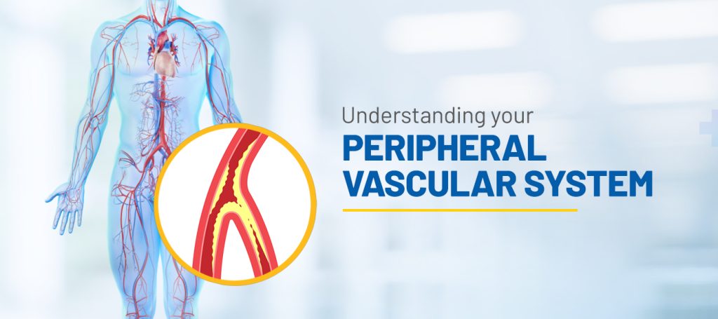
Peripheral Vascular Disease is the condition of blockage and narrowing of the arteries and veins in the legs, arms, and body parts other than the heart and the brain. Peripheral Artery Disease (PAD) and Peripheral Vascular Disease (PVD) are often used interchangeably. However, there is a difference between the two. PAD is the narrowing of the arteries due to the accumulation of plaque (i.e., fats and cholesterol) on the inner walls of the arteries that impair the blood flow from the heart to different parts of body, which remain deprived of oxygen and nutrients. While PVD refers to the blockage and narrowing of the arteries and veins in parts of the body other than the heart and the brain due to various reasons, including the accumulation of plaque. Thus, PVD is an umbrella term.

Causes of PVD

The primary cause of PVD is Atherosclerosis. The build-up of plaque, i.e., accumulation of fats and cholesterol, can be in any artery in the body. The fatty substances and cholesterol that block the arteries and thereby reduce the blood flow are known as Atheroma. The condition of this plaque formation in the arteries is called Atherosclerosis. Due to narrowing, when the arteries cannot supply enough blood to the legs, it causes pain, cramps, aches, and discomfort during physical activity, but the same subsides with rest. This condition of pain in the legs is called claudication. Atherosclerosis can happen in any body part. But it may be noted that Atherosclerosis is caused in the arteries and not in the veins.   

Besides, Atherosclerosis, the other responsible factors for PVD can be-

  • Coronary Artery Disease – people with blocked arteries of the heart are at risk of developing PVD.
  • Lifestyle – Lifestyle choices like smoking, sedentary living, poor eating habits, and drug use also play a significant role in developing PVD.
  • Medical conditions– Medical conditions include diabetes, high blood pressure, high cholesterol, obesity, and kidney disease, which may also be the reason for one developing PVD.
  • Injury – Injury to the legs and arms may cause the veins to narrow or swell, impairing blood flow from the legs or arms to the heart.

Symptoms of PVD

People with PVD may not experience any symptoms in the early stages. However, the symptoms depend on the blood vessels blocked and the extent to which they are blocked. Some of the symptoms that people affected with PVD may experience are –

  • Pain with physical activity, i.e., cramps, aches in the hip, buttocks, calf, and thigh which subside with rest.
  • Weakness in muscles
  • Shiny and smooth skin
  • Swelling, sores in legs that do not heal
  • Numbness and coldness in toes, feet
  • Reduction of pulses in feet
  • Decolouration of the skin
  • Severe burning
  • Loss of hair in the affected area

Types of PVD

Depending upon the nature of damage caused to the blood vessels, PVD is categorized as Occlusive and Functional.

  • Occlusive PVD

When there is physical damage to the vessels, resulting in structural changes in the blood vessels, it is the case of occlusive PVD. The following are examples of occlusive PVD-

Atherosclerosis– When the walls of the arteries thicken or harden over a period due to the accumulation of plaque, obstructing the smooth flow of blood in the arteries.

Carotid Artery Disease– When the large arteries (carotid arteries) on either side of the neck carrying oxygenated blood to the brain are narrowed. It is known as Carotid Artery disease or Carotid Stenosis. 

Lymphedema– Lymphedema is a condition where the fluid in the lymphatic system builds up. The lymphatic system is the network of thin tubes and lymph nodes (small structures that filter harmful substances) running throughout the body to help eliminate body toxins and waste, fight against infection, and fuel the immune system. The body parts of the affected lymph nodes get swollen.

Abdominal Aortic Aneurysm – The part of the Aorta (the largest artery supplying oxygen-rich blood to all the body parts) that runs through the abdomen is called the abdominal Aorta. Aneurysm refers to the outer bulging (like a balloon or a bubble) on the blood vessel. This is caused due to weakening of the artery walls. An aneurysm may occur on the abdominal Aorta, which can be life-threatening with internal bleeding if there is a rupture in the artery. 

Deep Vein Thrombosis (DVT) – DVT is when a blood clot is formed in the deep vein, often in the leg, arm, or pelvis, resulting in swelling in the veins, cramping, and redness in the affected area. If the clot breaks, its fragments can travel to any body part.

Buerger’s Disease– This is chronic inflammation in the small arteries of the extremities (the hands or the feet), causing a blood clot and blockages in the legs or arms.

Popliteal Artery Entrapment Syndrome (PAES) – PAES is a rare vascular condition where the muscles and tendons near the knee compress the popliteal artery (the main artery behind the knee), thereby restricting blood flow to the lower part of the legs. This usually is common among athletes.

A few other diseases caused due to blockage of arteries leading to the particular organ of the body are Intestinal Ischemic Syndrome which affects the gastrointestinal system, and RenalArtery Diseasecausing damage to the arteries supplying blood to kidneys .The term Ischemic refers to the lack of blood supply to a body part that deprives it of oxygen and nutrients.

  • Functional PVD

When factors such as stress, cold atmosphere, smoking, or vibrating of machinery the blood vessels to widen or narrow, it is the case of Functional PVD. There is no physical damage or structural change in the blood vessels here.

Raynaud’s Disease– Exposure to cold temperature, anxiety, and emotional disturbances cause Raynaud’s disease, in which the blood flow to the fingers, toes, ears, etc., is impaired.

Chronic Venous Insufficiency (CVI) – CVI is a condition when the valves of the veins do not function properly and hence cannot transport blood back to the heart. The blood flows back to the veins instead of the heart, and this causes swelling or aching in the legs or feet. Varicose veins are twisted and enlarged veins visible under the skin caused by venous insufficiency.

Treatment of PVD

PVD affects cardiovascular health, and there is a high risk of life-threatening complications of heart attack and stroke if left untreated.

Healthy living with positive lifestyle changes like adequate physical activity, a proper nourishing diet low in fats, and smoking cessation can help avoid and reduce the risks of PVD.  

Timely treatment is of utmost importance in reducing the severity of PVD. The treatment starts with your doctor recommending various tests like-

  • Ankle Brachial Test, which measures the blood pressure at the ankle or the arms when the patient is at rest, or
  • A treadmill test is conducted to check the pain threshold while the patient walks on the treadmill.
  • Ultrasound Doppler to check the blood circulation in the arteries of the legs to ensure the presence of any blockage therein. 
  • Computed tomography angiography (CTA) and Magnetic resonance angiography (MRA) tests are carried out to detect clogged/narrowed blood vessels. 
  • Angiography- Angiography is conducted with the help of dyes that are injected into the arteries to detect the blockages.

Depending on the need and the severity of the condition, your doctor would suggest medications only or an interventional procedure with medications to treat PVD. 

Peripheral Angioplasty is a minimally invasive procedure performed under local anesthesia to open the clogged artery and restore normal blood flow. A diagnostic catheter (a thin tube) is inserted through a tiny incision in the thigh. With the help of a Live X-ray, the operator can guide this catheter to the site of the blockage in the artery. A balloon is inflated inside the artery to press the plaque to the walls of the artery, making way for normal blood flow. A stent support may also be provided to the treated artery to keep it open. Once the stent is placed correctly, the incision is closed.

Conclusion

PVD is a slow and gradually increasing circulatory disorder. The symptoms are not noticeable at the early stages. However, those experiencing the symptoms must consult a doctor for timely and proper diagnosis and treatment. This is so because the major risks associated with PVD are heart attack or stroke complications which can be life-threatening as already mentioned above. In severe cases, this may also result in tissue death leading to limb amputation

Always follow a healthy lifestyle, and ensure timely and appropriate medical intervention and treatment to avoid and reduce the risks of Peripheral Vascular Diseases.

Knee replacement surgery is a common and effective way to restore mobility and relieve pain in patients with degenerative arthritis of the knee. In most cases, knee replacement surgery is successful and produces excellent results. The procedure restores much of the original functionality of your knee joint while also addressing any underlying causes of pain and disability. However, the very idea of having to undergo surgery creates stress and anxiety in many people, especially the elderly, which is quite normal and understandable. In such an event, immediate family members and caregivers need to step in and do what it takes to prepare them for the upcoming surgery. 

What is a knee replacement?

A knee replacement is a procedure in which a surgeon replaces your knee joint with an artificial joint. The artificial joint is made of a metal and plastic component that is surgically implanted. Knee replacement surgery treats people who have advanced osteoarthritis or rheumatoid arthritis. Knee replacement surgery restores function to the knee joint. You may notice an increased range of motion and decreased pain after knee replacement surgery. 

How to Prepare Seniors for Knee Replacement Surgery

Preparing our elderly folks for surgery is one big factor in the success of the surgery. The surgery’s run-up is critical to success, healing, and recovery. Knee replacement surgery is a major procedure that can significantly improve mobility and reduce pain for elderly individuals with severe knee damage or degeneration. Preparing for knee replacement surgery can be a challenging process, especially for elderly individuals who may have additional health concerns or mobility issues. 

The role of caretakers includes:

  • Taking care of the patient’s emotional health.
  • Sharing success stories of other patients to boost their morale.
  • Ensuring their good health and diet before surgery.
  • Providing moral support.
  • Creating an environment of acceptance and readiness to help them come through successfully.

Here are some steps you can take to help prepare your elderly loved one for knee replacement surgery:

  • Help them understand the surgery: Make sure they understand the surgery well and what to expect before, during, and after the procedure. Of course, the surgeon and their team will brief the patient and their immediate family about the surgical procedure, what it entails and the outcome, and all the nitty-gritty. However, as a caregiver, it is your responsibility to try and comprehend the medical implications of this surgery and pass on the net positive information to the patient. This hand-holding will also reduce the fear, stress, and nervousness that come before undergoing a surgical procedure. 
  •  Follow the surgeon’s instructions: The surgeon will provide specific instructions on preparing for the surgery, such as fasting for a certain period before the procedure. It’s also important for the elderly to follow their surgeon’s instructions and attend any pre-surgery appointments or educational sessions. By preparing in advance and working closely with their healthcare team, they can increase their chances of a successful surgery and recovery.
  • Discuss medications and allergies: Make sure to inform the surgeon and anesthesiologist about any medications your loved one is currently taking and any allergies they may have.
  • Do Your Research: Additionally, reading up on doctor-backed articles and information about the procedure for the layperson is helpful. Do make sure all doubts and questions are addressed before the surgery. Constant reassurance to your elderly loved one that you will be there before, during, and after the surgery. 
  • Attend pre-surgery tasks and appointments: Your loved one must attend all pre-surgery appointments with their surgeon and anesthesiologist to discuss the procedure and any potential risks or complications. Help them gather necessary medical documents or reports, fill out paperwork, and arrange transportation to and from the hospital.
  • Prepare the home: Consider making any necessary adjustments to the home to make it easier for your loved one to move around after the surgery and make the recovery process more manageable. This may include installing handrails, removing tripping hazards, and ensuring all necessary items are within easy reach. This assures them that their needs and comfort are taken care of and acts as a morale booster.
  • Help them get in shape: Encourage them to engage in gentle exercises to strengthen their muscles and improve their overall physical condition before surgery.
  • Make arrangements for aftercare and recovery plan in place: Assure your elderly loved ones that post-surgery, there will be someone to help them with daily activities such as bathing, dressing, and arrange for their healthy meals, balanced diet, medications, and other routine needs.
  • Keep Expectations Realistic: Any knee surgery has its set of ups and downs in terms of absolutely no 100 percent guarantee despite both physical and mental preparations. The patient’s anxiety cannot be fathomed. It is in our hands how we handle questions around the tenure of recovery, the physical limitations during the recovery process, and what recovery and rehabilitation look like. 
  • Be Prepared About The Risks: Both the patient and the caregiver must be ready and prepared for the risks of such surgeries. They include infection around the surgical site, damage to blood vessels that can lead to gangrene or even limb amputation, breathing trouble due to anaesthesia, damage to nerves, or metal allergies from the metals used in the artificial joints. 
  • Keep their spirits up: Surgery can be stressful, so it’s important to offer emotional support and encouragement to help them stay positive and focused. They will feel irritable and anxious because of their immobility. They will feel tremendous helplessness due to their dependence on others. There will be bouts of low self-esteem and lack of confidence as well. Offer emotional support and reassurance to help them feel more comfortable and at ease.

Knee replacement surgery is a common and effective way to restore mobility and relieve pain in knee osteoarthritis patients. Many people dread getting a new knee, while others may have reservations. However, helping elderly patients know what knee replacement surgery entails and preparing them for it can make the process less stressful with their positive acceptance and mindset. 

What should I not do before knee replacement surgery?

 Avoid alcohol, tobacco, and nonsteroidal anti-inflammatory drugs to prevent surgical blood loss. 

How long does it take a 70-year-old to recover from knee surgery?

– It takes about 3-6 months for a full recovery.

How long do you need someone with you after knee replacement surgery?

 Post-operation, a patient needs a caregiver for 2-3 weeks for support and exercise assistance till they gain confidence and independence from pain.

What do you need at home after knee surgery?

Lots of care and support besides good nutrition, timely medications, and exercise assistance.

Introduction

Aortic aneurysm, as the name suggests, happens in the aorta. It is typically linked with the hardening of the arteries, also known as atherosclerosis. This condition may be inherited or a result of complications due to high blood pressure or smoking. Aortic aneurysms start as small bulges which eventually grow in size as the flow of blood puts continuous pressure on the wall of the aorta, the major blood vessel that carries blood from the heart to the rest of the body. They occur when part of a blood vessel’s wall weakens and gives way to the bulge, typically the abdominal aorta. A tear in this weakened area allows blood to escape from it. This is known as an aortic dissection. If the aneurysm occurs in the chest region it is called in Thoracic Aortic Aneurysm (TAA), if it occurs in the abdominal region, it is called Abdominal Aortic Aneurysm (AAA). The most common site in the abdomen is below the area where the aorta gives out branches to supply blood to kidneys, it is called an Infrarenal abdominal aortic aneurysm. But they can also occur in the thoracic region, where there are fewer blood vessels. Aneurysm can be associated with other conditions such as Marfan syndrome, and Ehlers-Danlos Syndrome, hypertension and trauma. It also occurs along with heart valve disease likes bicuspid aortic valve stenosis.  

What Causes Aortic Aneurysm?

A weakening of the aorta or aortic wall causes an aortic aneurysm. The most common causes are atherosclerosis and high blood pressure. This happens due to a combination of factors such as ageing, certain genetic disorders, and certain drugs. This can be present at birth or can develop as a result of disease or injury. Aortic aneurysm is not the same as aortic stenosis, which is when there is constriction of the aorta due to the narrowing of the aortic wall.

Types of Aortic Aneurysms

Aortic aneurysms are classified by their size into small, medium, and large categories. The most severe threat of an aneurysm is that it can burst and cause a stroke or massive bleeding, which in either case is life-threatening. A large aneurysm can affect blood circulation leading to blood clots.

Causes of Aortic Aneurysms

The most common causes are

Atherosclerosis- Accumulation of cholesterol in the artery can lead to degeneration of its wall on enlargement. This condition is called atherosclerosis and is the most common cause of aneurysms.

 High Blood Pressure- High Blood pressure puts stress on the wall of aorta. Over the years this stress can lead to bulging of the blood vessel which may be the leading factor for an aneurysm.

Diabetes- Uncontrolled diabetes damages blood vessels by accelerating the process of atherosclerosis, in turn leading to an aneurysm.

Injury / Trauma- An injury to the chest or abdomen can damage the area of the aorta leaving it vulnerable to bulging.

In many cases the cause of aneurysm goes unknown.

Risk Factors involved

Most aneurysms occur in people aged 55 years or older. Although thoracic aneurysms and dissections are known to occur in younger individuals also. Aneurysms are more common in men than women. High blood pressure, smoking and family history.

Symptoms

The symptoms can range from mild to severe, sometimes none at all. But some people are at higher risk of getting one, complete physical examination can help early detection of warning signs.

It’s important to see your doctor right away if you experience any of the following symptoms:

  • Sudden and tearing pain
  • Chest Pain- Often radiating to the back and shoulders.
  • Back Pain – Often mistaken as spine problems.
  • Abdominal Pain
  • Shortness of breath
  • Hoarseness of voice
  • Hemoptysis (coughing up blood)
  • Hemotemesis (Bloody Vomits)
  • Lump near a blood vessel
  • a new bulge in your abdomen, or
  • a sudden change in your heart rate.
  • Aortic regurgitation, when the regurgitation of blood from the aorta causes backflow into the chest. This symptom can be mild or severe.
  • Aneurysm rupture can cause a person’s abdomen to swell causing severe abdominal pain and tenderness which is life threaten and requires immediate emergency care.

Diagnosis

Aneurysm is generally detected by an echocardiogram that can reveal the size and shape of the aorta and if an aneurysm is present. Blood tests can also show an increase in certain proteins that can be present if the aneurysm is large enough to cause blood loss. In a medical diagnosis, your doctor will ask questions on the following,

  • If there is a family history of heart disease
  • Complete physical examination of checking your blood pressure, feel the abdomen for a lump/mass
  • If your doctor suspects an aneurysm, you will be asked to get an ultrasound / echocardiogram / chest x-ray / MR Angiography / CT / Trans esophageal  echo test depending upon the location of aneurysm.

The doctor can also recommend to get a minimally invasive test done known as angiogram.

Treatment of Severe Aortic Aneurysms

Since rupturing of aneurysm is life threatening, it is necessary to get immediate medical attention. Treatment is through medicines or surgery/intervention depending upon site, size and growth rate of aneurysm. Routine testing is recommended to ensure that the aneurysm is not growing. Surgery / Intervention is advised if the aneurysm is growing quickly to avoid a rupture and complications. Aneurysms are unlikely to rupture if they grow slowly and are less than 5.5 cm (about 2 inch) in diameter and causing no symptoms. Serial Ultrasound / CT Scan every 6-12 months to monitor the aneurysm size is the proper treatment for most people along with blood pressure control medicines.

An aneurysm larger than 5 to 5.5 cm (2 inch) requires intervention. Aneurysms that are enlarging rapidly causing symptoms or showing signs of a probable rupture (such as leaking) require immediate medical intervention. Delaying this put an individual at even greater risk of rupture and dead. Newer treatment modalities like endovascular stenting are available to treat large aneurysms using percutaneous techniques avoiding surgery. Stenting is also now providing to be useful in rupture / leaking in advanced centers

The aneurysm repair aims to strengthen the aortic wall to prevent it from weakening again. An aneurysm repair is usually done under general anesthesia.

Prognosis of Severe Aortic Aneurysms

Generally, people with small aneurysms are not expected to have problems. Most people with aortic aneurysms are expected to have a normal lifespan and not experience any problems with their aorta. However, 10% to 15% of people will have an aneurysm that is large enough to cause significant symptoms, like significant pain or severe swelling. And 1% of people will have a rupture of the aneurysm.

Conclusion

As you now know, an aortic aneurysm is a condition in which part of your aorta – the major blood vessel that carries blood from your heart to your body – becomes enlarged. Call the doctor immediately if you think you may have an aneurysm because if that ruptures, it can be life-threatening. At best, to prevent an aortic aneurysm, you can control your blood pressure, and should you have a family history of heart disease or stroke, then bring changes to improve your health and lifestyle by exercising regularly, watching what you eat, and completely stopping smoking.

FAQS

What are the early symptoms of aortic aneurysm?

Early symptoms of aortic aneurysm include a pulsating feeling in your stomach and nagging back pain.

How serious is an aortic aneurysm?

The aorta is the largest blood vessel in the body which runs from the heart through the center of the chest and abdomen. Therefore, a ruptured abdominal aortic aneurysm can be life-threatening.

Can a person survive an aortic aneurysm?

Some aortic aneurysms are hereditary or congenital, such as bicuspid aortic valve, infection, or inflammatory conditions. You can live with an aortic aneurysm. There are many ways to prevent dissection or a rupture with periodic examinations, medications, and healthy lifestyle.

What happens when you have an aortic aneurysm?

When you have an aortic aneurysm, there will be a bulge or swelling in the aorta, the main blood vessel that runs from the heart down through the chest and tummy. This can get bigger over time and could burst (rupture), can be life-threatening.

What should you not do with an aortic aneurysm?

Your doctor will advise you to avoid heavy lifting and intense physical activities, which can increase blood pressure, putting additional pressure on your aneurysm.

Can you fully recover from an aortic aneurysm?

Typically, you will need 2 to 3 months to fully recover from an aortic aneurysm and be able to resume many of your usual activities after 4 to 6 weeks post treatment.

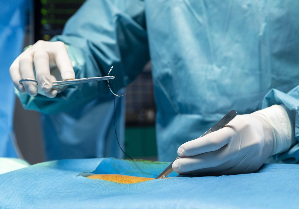
Suture and Needle choices vary depending on the type of surgery and the patient’s needs. Therefore, knowing the differences between these different types of sutures and when to use them is important to get the best outcome from your surgical procedure. Using the wrong type of suture can lead to undesired surgical outcomes, damage to surrounding tissue or organs, and chronic pain. This can lead to undesired wound healing and complications during recovery, which may require re- suturing for proper wound healing. Suture usage vary on the type of Tissue, patient age as well as the Type of surgical procedure in which suturing needs to be performed. Suturing promotes wound healing by approximating the layers of the tissue and enabling the natural healing process. It also closes the wound thus avoiding the wound to get contaminated by the atmosphere.

What is a suture?

A suture is a surgical device used to close any surgical or traumatic wound requiring skin & tissue closure. A closure is intended to primarily repair skin and deeper layers of the tissue and fascia to fast-track wound healing. Some examples of sutures include monofilament sutures, monofilament polyester sutures, and large absorbable sutures. They are used for repairing cuts and wounds that are deep and open. Sutures are used to close wounds by first suturing the edges together and then using the suture to tie off the wound. After the wound has healed, the suture is either removed or absorbed naturally into the body.

Types of Sutures

Historically, similar to modern sutures, different forms of wound closure mechanisms were employed with needles made of bone and suture materials running from flax, bark, silk, human hair, reeds, etc. Today, there are many options for sutures. Hence, it becomes crucial to make the right suture choice, which means understanding the purpose, attributes, and features of each type of suture. It also means understanding the difference in filament types and the different needle types and under which clinical procedures they have been designed for use. The correct suture helps the surgeon with the right technique and choice of incision to execute and handle the surgical procedure, provide the correct strength, and secure the knots in a manner that can handle and tolerate wound changes during the healing process.

Suture material types are classified as follows:

Absorbable and Non-absorbable Sutures

Absorbable sutures, which get absorbed by the body naturally, are typically employed for deep temporary closure for tissues to heal, and the sutures lose most of their tensile strength over a period of time ranging from weeks to months. Non-absorbable sutures do not get absorbed by the body and hence are required to be removed after a few days of the surgery or may be left in the body permanently. These sutures are used for long-term tissue closures, wounds that take time to heal, like in the case of hernia fascia defects closure. 

Natural and Synthetic Sutures

Sutures are further classified into natural and synthetic sutures. Natural sutures are derived from natural substances. For example Catgut Sutures are usually derived from collagen derived from bovine Guts (used for internal lacerations or wounds). Another Examples of Natural sutures are silk sutures (used in general soft tissue closure and ophthalmic surgery). Catgut degrades by enzymatic reactions (breaking down of proteins partially as peptides or completely as amino acids). They can cause inflammation at the suture site. Synthetic sutures are derived from artificial sources and are made from materials like Nylon sutures (used for general closures and plastic surgery) which is non-absorbable in nature. Synthetic absorbable sutures like Polydioxanone (PDS) sutures (used in abdominal closures or pediatric cardiac procedures) degrade by hydrolysis (the polymer structure of the filaments gets broken down with water penetrating the suture strands) which causes less inflammation at the surgical site. 

Monofilament or Multifilament

Based on the structure, sutures are classified as Monofilament or Multifilament sutures.

Monofilament sutures are single filament sutures with less surface area and higher memory. By memory, we mean the tendency and capacity of a suture to return to its original shape, which makes a suture significantly prone to the knots loosening. They require greater handling and care in the form of more knots to ensure security and less fracture. Monofilament sutures have the capacity to pass through tissues easily, causing less inflammation. 

Whereas Multifilament sutures, as the name suggests, are braided or twisted sutures that are easier to handle by the surgeon as they hold the knots with greater security and less memory. They are prone to inflammation and infection. Multifilament sutures are more expensive than traditional sutures. 

The following are the most commonly used sutures in Surgeries: 

Monofilament sutures, made of a single synthetic strand, are typically used in skin

Closures and Ligation of blood vessels. 

Polypropylene sutures are the synthetic non absorbable sutures; these are typically used in soft tissue closures and are less likely to cause allergic reactions.

Dressings sutures are used to close wounds and stop bleeding. They are typically made of silk, Nylon, or synthetic materials. 

Epithelial sutures are used to close and repair minor wounds. They are made of proteins and are typically used with tissue types such as skin, mucous membranes, and cartilage.

Soft Tissue sutures are used to close wounds in soft tissue such as muscle and tendons. They are also used to support joints and may be made of polyester or polypropylene.

Some sutures are specially used to repair veins and arteries. They are made of polypropylene, Polyester & Nylon.

Needle Types 

The needle is indeed the next most important aspect of sutures after identifying the correct suture type. Needle quality and the appropriate needle choice for a given surgery go a long way to determining the success of the surgery. The surgical needle is mainly built from stainless steel, making it corrosion-resistant. In surgical parlance, the needle has three main parts – the Swage point, the body, and the point. The Swage point is where the surgical suture attaches to the needle by stringing through. The body connects the eye to the point and decides the shape of the needle. In most cases, the needle is curved, but in some cases, it is straight. Curved needles come in different circle lengths like ¼, ½, ⅓, or ⅜ of a circle. The nature of the wound or tissue helps a surgeon choose the needle curve length. The surgeon needs to know where the needle tip is at all times, as it aids in skin closures with minimum trauma. A wrong surgical needle type can lead to tissue injury. The ideal needle is one that is flexible enough to bend yet rigid enough to withstand distortion. It should be sharp to pierce through tough tissues yet thin to cause minimum trauma. Stability of the needle to ensure accurate placement is also equally required. Thusan appropriate surgical needle must be sterile, corrosion-resistant, and firm enough to prevent any bending or distortion, prevent tissue trauma and injury, and ensure easy penetration. 

Different types of needles are based on the tip, like taper needles or cutting ones. Cutting needles have a tip with three sharp edges. In a typical conventional cutting needle, the cutting surface is inside the needle. In contrast, a reverse cutting needle used most commonly for sewing skin has it on the outside. Whereas taper needles are round and can be either blunt or sharp and are recommended for soft and delicate tissues and tendon repair. They typically pierce the skin without cutting it and spread the tissue as it passes through.

Summary

The right type of suture will prevent issues with excessive bleeding, damage to surrounding tissue or organs, and chronic pain. The thickness of the tissue, the location of the tissue, the risk of infection, and the amount of tension, along with different anesthesia requirements and patient age, play an important role in determining which type of suture to use. There needs to be more than the right type of suture to yield the desired surgical results. The correct choice of needle is equally essential and needed for a wound to be sutured with accuracy, precision, and minimal trauma, ensuring fast healing and preventing tissue reaction and inflammation.

FAQs :

Why is it important to use the correct suture technique?

It is important to use the correct suture technique for precision, minimal trauma to the wound at the surgical site and for faster healing.

Why is knowledge of suture material so important?

The knowledge of the right suture material is very important to the surgeon to maximize tissue healing and ensure scar aesthetics and minimal tissue trauma and inflammation at the surgical site.

What factors influence the surgeon’s choice of suture materials?

The main factors that influence a surgeon’s choice of suture materials are

  • Healing time of the wound or tissue
  • Wound type and strength
  • Properties of the suture material
  • Faster attachment of suture and tissue