Category

Surgery:

Category

Coronary Artery Disease (CAD) is the number one cause of death, disability, and human suffering globally. Once you are diagnosed with CAD you have to learn to live with it by adopting a lifestyle that fits you and your heart health. By lowering the risk factors, you can live your life despite having CAD.

There is a possibility that you may be living with CAD long before you realize it. It creates fear and anxiety due to the close association with heart attacks.

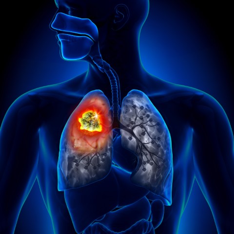
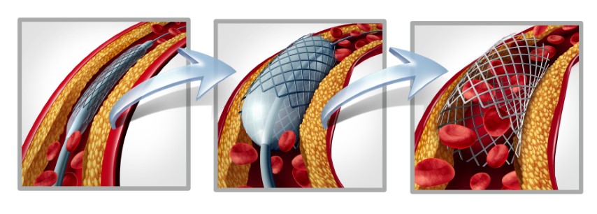
Coronary artery disease occurs when your heart’s primary blood arteries become damaged or diseased. Coronary artery disease is caused by cholesterol-containing deposits (plaques) in your coronary arteries and inflammation. The coronary arteries supply your heart with blood, oxygen, and nourishment. Plaque development can restrict these arteries, reducing blood flow to the heart. Reduced blood flow may eventually result in chest pain (angina), shortness of breath, or other signs and symptoms of coronary artery disease. A heart attack might be caused by a total blockage. Although Coronary Artery Disease (CAD) is not completely curable, the doctor would suggest an option of a procedure known as Percutaneous Coronary Intervention (PCI), which allows one to live a normal life. Coronary angioplasty, also known as percutaneous coronary intervention, is a non-surgical procedure for treating obstructive coronary artery disease that involves inserting a stent through a catheter (a thin flexible tube) into the blocked arteries.

Why is it done?

Coronary Angioplasty restores blood flow to the heart muscle and can improve symptoms of blocked arteries, such as chest pain and shortness of breath.

Does stenting improve long-term survival?

Not guaranteed. It saves your coronary, but stents do not increase a cardiac patient’s long-term survival rate. However, they do give a considerable early and sustained reduction in the requirement for subsequent treatments to reopen the treated artery. While studies have indicated that placing stents in newly re-opened coronary arteries reduces the need for repeat angioplasty procedures, it has also shown that stents do not affect death overtime. The findings have significant economic and clinical consequences for doctors considering whether to perform coronary artery bypass surgery or less-invasive angioplasty with stent implantation on their heart patients.5 According to medical opinion, your stents can help you live longer if you manage your other risk factors as per your cardiologist’s recommendations. Diet and exercise, however, are the most important factors to consider. Your risk factors for a heart attack, such as hypertension, diabetes, and obesity, can be controlled by the type and amount of food you eat. Along with these two critical components, follow your cardiologist’s advice and take your prescribed medicines on time to manage your blood pressure, diabetes, and cholesterol. As a result, even after implanting stents in three major coronary arteries, if correctly controlled, you can live a long life.

If you have a heart attack in your late thirties and have stents, is it likely you will not have an average life expectancy?

Over the last few decades, aging has been identified as one of the leading causes of heart attacks, affecting men and women aged 50 and up. People in their 20s, 30s and 40s are now more likely to suffer from cardiovascular attacks.

The guidelines emphasize lifestyle changes and the proper use of medicines as first-line treatment in adults with stable CAD. However, for people suffering from Non-ST Segment Elevation Myocardial Infarction aka NSTEMI (substernal pain while resting or with minimal exertion) and unstable angina; clinical insight is required to assess if other procedures such as CABG (Coronary artery bypass graft) or OMT (Optimal medical therapy)  are more appropriate.

Whatsoever the application, PCI should not be viewed as a ‘fast cure’ but rather as a procedure that should be discussed with your doctor to balance the benefits, risks, and limitations.

Source:

  1. https://www.narayanahealth.org/blog/coronary-artery-disease-life-expectancy-and-prognosis/
  2. https://www.mayoclinic.org/diseases-conditions/coronary-artery-disease/symptoms-causes/syc-20350613
  3. https://www.heartandstroke.ca/heart-disease/treatments/surgery-and-other-procedures/percutaneous-coronary-intervention
  4. https://www.onhealth.com/content/1/stents_save_coronaries_not_lives 
  5. https://www.verywellhealth.com/do-angioplasty-and-stents-prolong-life-4021221

The prostate gland is a small gland found only in males. It produces the seminal fluid that helps transport the sperms. The prostate gland is located below the bladder and in front of the rectum. As the name itself suggests, prostate cancer begins at the prostate gland, when the cells grow and multiply uncontrollably.

Prostate cancer is one of the most common types of cancer, and it may show no signs or symptoms in the early stages. However, patients with prostate cancer have higher chances of complete recovery if the cancer is detected early.

Symptoms of Prostate Cancer

As mentioned above, prostate cancer does not show any signs or symptoms during the earlier stages. Some common symptoms visible during the advanced stage of prostate cancer are:

  • Trouble urinating, or the need to urinate more often than usual
  • Blood present in the urine
  • Blood in the semen
  • Erectile dysfunction
  • Pain in the bones – especially the hips, spine, and ribs
  • A feeling of weakness/numbness in the feet
  • Loss of weight

While most of these signs and symptoms may point to several other underlying conditions or diseases, it is necessary to talk to your healthcare provider if these symptoms are persistent or continue to worsen.

What causes prostate cancer?

Though the causes of prostate cancer remain unclear, it is known prostate cancer occurs when the cells in the prostate mutate. The mutation causes cells to grow and multiply rapidly, forming abnormal cells. The abnormal cells continue living, while the healthy cells die. These growing abnormal cells form a tumor and invade the nearby tissues or organs. Gradually, these abnormal cells also spread to the other parts of the body, causing cancer to grow.

Risk Factors of Prostate Cancer

Several factors can increase the risk of prostate cancer, such as:

Age: The risk of prostate cancer increases with age. Prostate cancer is more prevalent among men over the age of 50.

Ethnicity: For reasons unknown and undetermined, prostate cancer is more common among men of African-American descent. As per to urology.org, African-American men tend to get diagnosed with prostate cancer at an earlier age, and the cancer is likely to be aggressive in growth.

Family History: Men with a family history of prostate cancer have higher chances of being diagnosed with the same. A strong family history of breast cancer can also increase the chances of one developing prostate cancer.

Obesity: People who are obese have higher chances of developing prostate cancer, compared to those who maintain a healthy weight.

Prostate Cancer Diagnosis

If your doctor suspects that you might have prostate cancer, he might enquire about your symptoms such as urinary and sexual problems. He might also ask you about possible risk factors such as your family history to analyze the chances of you developing prostate cancer. after this, the doctor will run several diagnostic tests to diagnose the disease:

Digital Rectal Exam (DRE): During this, the doctor might insert a gloved finger into your rectum to examine the prostate, and feel for any bumps or hardened areas. If the doctor finds any abnormalities in the shape or size of the gland, he may advise further tests.

PSA Blood Test: A blood sample is taken and tested for PSA. PSA stands for Prostate-specific Antigen, which is a protein produced by the prostate gland. It is normal for a small amount of PSA to be found in your bloodstream. However, if it is higher than usual, it may be an indication of prostate cancer.

Ultrasound: During this test, a small probe is inserted into the rectum, and uses soundwaves to create images of the prostate gland.

MRI: Magnetic Resonance Imaging (MRI) scan may be suggested by your doctor to create a more detailed picture of your prostate gland.

Biopsy: The doctor may also collect a sample of cells in your prostate gland to check for signs of cancerous cells. This procedure is known as a prostate biopsy. A thin needle is inserted into the prostate to collect a sample of the tissue.

If you are diagnosed with prostate cancer, the doctor may use tests such as ultrasound, CT scan, or MRI to determine the stage of cancer.

Treating Prostate Cancer

The treatment for prostate cancer depends on how aggressive the cancer is and the extent it has spread. The right treatment option will also be chosen depending on the side effects and their impact on overall health.

Surgery: This involves removing the prostate gland, along with some nearby tissues and lymph nodes. Surgery is usually performed when the cancer is confined to the prostate. It may also be used to treat advanced prostate cancer combined with other treatment methods.

Radiation Therapy: This involves using high-powered radiation to kill the cancerous cells. This can be performed externally (external beam radiation) or internally (brachytherapy), by placing radiation sources in the prostate tissue.

Cryotherapy: This treatment involves using extremely cold gases to freeze the prostate tissue. After this, the tissue is allowed to thaw, and the procedure is repeated.

Hormone Therapy: This treatment stops the body from producing testosterone, the male hormone. Since prostate cancer cells rely on testosterone to grow, cutting the hormone supply may cause the cancerous cells to die.

Chemotherapy: This therapy uses drugs to kill the rapidly growing cancerous cells. Chemotherapy is usually used when cancer has spread to the other organs and areas of the body.

Immunotherapy: As the name itself suggests, this therapy involves using the immune system to fight cancer cells. The immune system usually does not attack cancer cells as they produce proteins that help protect the cancer cells. Immunotherapy interferes with this process.

Targeted Therapy: This treatment emphasizes the abnormalities present in the cancerous cells. Targeted therapy blocks and combats these abnormalities, causing the cancer cells to die.

Though several factors increase the risk of developing prostate cancer, early detection and following a healthy lifestyle can help nip prostate cancer in the bud. Though certain risks such as age, family history, and ethnicity cannot be avoided, other factors such as obesity can be avoided by maintaining a healthy diet and exercising regularly. Talk to your healthcare provider immediately if you notice certain symptoms and take the necessary precautions!

Lung cancer is the most common cancer in the world. According to the World Health Organization, there were 2.21 million cases in the year 2020. It is the most common cause of death around the world. Patients have a 13 times better chance of living for five years if they are diagnosed early. Lung Cancer Awareness Month (LCAM) is celebrated every year in November to raise awareness about the disease and continue to challenge the stigma associated with lung cancer.

The goal of the month is to urge individuals to seek medical assistance sooner rather than later, to encourage early diagnosis so that patients have the best chance of a successful treatment, and to emphasise other key aspects that affect patient outcomes. Educating people on the complexity of lung cancer, the vast spectrum of people who are affected, and the harmful effects of lung cancer stigma can aid in earlier diagnosis and better patient treatment. Furthermore, LCAM is also an occasion to highlight therapeutic developments, advocate for global access to care, and, most importantly, demonstrate our support for patients and their loved ones.

Facing the stigma

Lung cancer is often misunderstood to be solely a smoker’s disease. However, more than half of individuals diagnosed are former smokers or non-smokers. This misunderstanding has been associated with poor outcomes due to factors like waiting too long to seek treatment, disease-related distress, a lack of social support, and poor care quality.

What you need to know about Lung cancer

Cancer is a condition in which the cells of the body grow out of control. Lung cancer is cancer that starts in the lungs and spreads throughout the body. Lung cancer begins in the lungs and can spread to the lymph nodes or other organs in the body, including the brain. Cancer that has spread to other organs may extend to the lungs as well. Metastases are the spread of cancer cells from one organ to another.

There are two types of lung cancer:

Small cell lung cancers (SCLC) and non-small cell lung cancers (NSCLC) are the two forms of lung cancer. This classification is based on the appearance of tumour cells under a microscope. Making the distinction between these two types of tumours is critical since they develop, spread, and are treated differently.

SCLC accounts for roughly 10% to 15% of all lung malignancies. This form of lung cancer is the most aggressive and fastest-growing of all. Cigarette smoking is highly linked to SCLC. SCLCs spread quickly throughout the body, and they are usually detected after they have spread widely.

The most common type of lung cancer is non-small cell lung cancer (NSCLC), which accounts for roughly 85% of all occurrences. NSCLC is divided into three categories based on the cells detected in the tumour. They are as follows:

  • Adenocarcinomas, like other lung cancers, are linked to smoking, this form is also seen in nonsmokers, particularly women, who get lung cancer. The majority of adenocarcinomas develop in the lungs’ periphery. They have a proclivity for spreading to lymph nodes and beyond.
  • Squamous cell carcinomas used to be more common than adenocarcinomas, but now they make up around 25% to 30% of all lung cancer cases. Squamous cell tumours are most common in the bronchi of the central chest. This type of lung cancer tends to stay in the lung, spread to lymph nodes, and grow large enough to produce a cavity.
  • Large cell carcinomas, also known as undifferentiated carcinomas, are the least prevalent kind of NSCLC, accounting for 10% to 15% of all lung cancer cases. This malignancy has a significant proclivity for spreading to lymph nodes and distant locations.

Types of Treatment

Depending on the type of lung cancer and how far it has gone, there are numerous treatment options. Surgery, chemotherapy, radiation therapy, targeted therapy, or a combination of these treatments can be used to treat non-small cell lung cancer. Small cell lung cancer is usually treated with a combination of radiation and chemotherapy.

  • Surgery: A procedure in which surgeons remove cancerous tissue from the body.
  • Chemotherapy: Special medications are used to shrink or kill cancer cells. The drugs can be pills that you consume or medicines that are injected into your veins, or both.
  • Radiation therapy: Treatment that involves the use of to kill cancer, high-energy rays (similar to X-rays) are used.
  • Target therapy: Drugs are used to stop cancer cells from growing and spreading. The drugs can be taken orally or administered intravenously. Before targeted therapy is employed, tests will be performed to determine if it is appropriate for your cancer type.

In the treatment of lung cancer, doctors from various specialties frequently collaborate. Pulmonologists are doctors who specialise in lung illnesses. Surgeons are medical professionals who carry out procedures. Thoracic surgeons specialise in surgery of the chest, heart, and lungs. Medical oncologists are doctors who specialise in using medications to treat cancer. Radiation oncologists are doctors who use radiation to treat cancer.

It is important to educate yourself and your loved ones about the most common type of cancer so that lives can be saved. If you notice anything unusual contact your doctor immediately.

Hemorrhoids are one of the most common anorectal disorders; according to NCBI it affects around 25% to 30% of the population. People of all ages, genders, races, and ethnicities are affected. Piles become increasingly common as people become older, affecting more than half of those over the age of 50. Hemorrhoids are very common, so don’t be shy to get help! Hemorrhoids are bulging, inflated veins that occur in the anus and rectum (Back passage). They can be unpleasant, painful, and cause rectal bleeding. Hemorrhoids, are natural cushions we’re all are born with, although they don’t bother us at first. They only cause uncomfortable symptoms when they become swollen and enlarged, then known as swollen piles.

Hemorrhoids (Piles) are a topic that most people avoid discussing and prefer being silent about. But being silent may prevent you from receiving treatment for a common ailment and which can land you up in severity. Your doctor isn’t shy about bringing up the subject of haemorrhoids, so discuss freely. 

When to see the doctor? 

Most people think of hemorrhoids as a minor problem, it can be very painful. It is important to know when to treat hemorrhoids on your own and when to seek help, as it can avoid unnecessary complications. 

Make an appointment with your doctor if you’re having any of the following symptoms related to your hemorrhoids:

  • You notice bright red blood on your toilet paper or have rectal bleeding.
  • Your rectum or anus is causing you agony and discomfort.
  • You’ve tried over-the-counter medications for more than a week and they haven’t helped you.
  • You experience a maroon or dark tar-colored bowel movement, which could be an indication of bleeding.
  • You feel mass outside your back passage

If your rectal bleeding won’t stop and you’re feeling dizzy or faint, it’s a medical emergency that necessitates a visit to the Emergency room (ER). 

 How do doctors treat Haemorrhoids? 

 Hemorrhoids are treated by doctors in the office, in an outpatient clinic, or a hospital. Treatment depends on severity. For an initial stage of piles lifestyle changes can help you if given at on right time. For mild piles office procedures can help you. But for severe piles surgery is the only option.

Office procedures for very mild piles:

  • Rubber band ligation: Rubber band ligation is a treatment that surgeons employ to treat internal hemorrhoids that are bleeding or prolapsing. A doctor wraps a specific type of rubber band around haemorrhoid’s base. The band stops the blood supply. Within a week, the banded component of hemorrhoid shrivels and slumps.
  • Sclerotherapy: In this therapy, a surgeon injects a special chemical into haemorrhoids, which causes the haemorrhoid to shrink, while also stopping it from bleeding.
  • Photocoagulation with infrared light: A doctor employs an infrared light instrument to treat internal hemorrhoids. Scar tissue forms as a result of the infrared light’s heat, cutting off the blood supply and reducing hemorrhoids.
  • Electrocoagulation: A doctor inserts an electric current into an internal hemorrhoid with an instrument. Scar tissue forms as a result of the electric current, cutting off the blood supply and reducing hemorrhoids.

Surgical procedures for moderate to severe piles:

  • Haemorrhoidectomy: A haemorrhoidectomy is a procedure in which a surgeon removes the prolapse of hemorrhoids (which is coming out from the back opening) by cutting it directly with a scalpel, scissors, or electrical energy source.
  • MIPH (Minimally invasive procedure for Hemorrhoid): Internal hemorrhoid tissue is removed and the prolapse of the internal hemorrhoid is pulled back into the anus by using a special stapling circular device.

Today, let us know more about MIPH 

MIPH (Minimally invasive procedure for Hemorrhoid) or Stapled haemorrhoidopexy is a surgical procedure for treating hemorrhoids. It is the preferred therapy for third and fourth-degree hemorrhoids (a severe form of piles that protrude with straining and are visible on physical examination outside the anal margin. The manual reduction must be done on a regular or irregular basis). Stapled haemorrhoidectomy is a misnomer because the procedure removes the unusually slack and enlarged tissue which supports hemorrhoids that have caused the hemorrhoids to prolapse downward, rather than the hemorrhoids themselves.

A circular, short hollow tube is introduced into the anal canal for stapled haemorrhoidopexy. A suture (a long thread) is woven circumferentially through the anal canal above the internal hemorrhoids through this tube. The stapler (a disposable instrument with a circular stapling device at the end) is inserted into the hollow tube, and the suture ends are brought together and pulled inside the stapler along with prolapsed hemorrhoids. Stapler when fired causes cutting of slack tissue and stapling of hemorrhoids back to their original position.

 Patients who undergo a minimally invasive procedure for hemorrhoids (MIPH)/ Stapled Haemorrhoidopexy benefit from the following:

  • Return to work as soon as possible
  • There is very little discomfort
  • There are no cuts, hence there are no dressings required
  • There are no follow-ups
  • Released from the hospital in a day

Comparison between MIPH and Conventional Haemorrhoidectomy

Haemorrhoidectomy is a procedure that removes hemorrhoids from the body. You will be given general or spinal anesthesia to prevent you from feeling discomfort. Around hemorrhoids, incisions are made in the tissue. To prevent bleeding, the enlarged vein inside the hemorrhoid is tied off, and the hemorrhoid is removed. It is possible to suture the surgical area to shut it or leave it exposed. The wound is covered with medicated gauze. A knife (scalpel), an electric tool (cautery pencil), or a laser can be used to do surgery. In most cases, the procedure is performed in a surgical center. You’ll probably return home the next day or more.

Stapler Haemorrhoidectomy / MIPH (Minimally Invasive Procedure for PILES) is a procedure where an expert surgeon employs stapling equipment to remove a portion of the pile mass with a staple gun, addressing two major concerns: bleeding and prolapsed hemorrhoids. The titanium staples not only cut but also seal at the same time, which lowers bleeding and post-operative pain. Because no extra incision on the lower region of the anal canal is necessary, the patient does not require any post-operative dressing. A patient can return home the same day and resume normal life in a day or two.

The most prevalent complaints of piles are painless rectum bleeding and haemorrhoidal prolapse, painful defecation. When compared to open haemorrhoidectomy, the average length of surgery for MIPH and post-operative bleeding is much shorter. The postoperative pain score and subsequent need for analgesics are significantly lower in the MIPH. MIPH patients have a faster average wound healing time and a quicker return to routine activities. Less recurrence or incontinence is observed. Similarly, there are no significant differences in long-term problems between MIPH and traditional open haemorrhoidectomy in patients with MIPH.

MIPH is a frequently used and safe procedure for grade III and IV hemorrhoids. It results in less pain, a shorter hospital stay, and fewer early postoperative problems. Wound healing and return to normal activities are faster, and long-term problems are not significantly different.

Knee replacement, also known as knee arthroplasty, is a surgical procedure done to reverse knee damage, to relieve pain and disability caused by conditions such as arthritis. For people suffering from severe knee pain caused by osteoarthritis, rheumatoid arthritis, or any other similar ailment, knee replacement surgery may be able to offer relief. Whether you need to undergo a surgical procedure or not is a decision that you and your doctor will collectively take.

The procedure for knee replacement involves cutting away the damaged bone and cartilage and replacing it with an artificial joint made of metal or plastic polymer.

The treatment methods for arthritis and other conditions as such are advancing day by day and providing better and more optimal long-term relief. This is where the futuristic robotic knee replacement surgery comes in. A lot of people have queries about the procedure and its after-effects. Here are 5 important things you should know about Robotic Knee Replacement Surgery.

  • A robot does not perform the surgery

The robotic technology’s state-of-the-art 3D software lets your surgeon design and plan the surgery much in advance, making necessary adjustments in real-time to provide a more precise alignment of the implant as compared to conventional non-robotic partial knee replacement.

While up to 20% of patients are not happy with the outcome following Knee Replacement surgery, the use of a robot as an additional surgical tool helps the surgeon during all stages of the knee replacement surgery and helps in delivering the best possible outcome.

  • Improved accuracy

Robotic assistance technology enhances the surgeon’s expertise, preventing accidental surrounding tissue damage aiding in greater precision during surgery. It helps the surgeon to pre-plan bone cuts and implant sizes, unique to your anatomy.

The 3D images of the knee obtained through a CT scan help the surgeon to specialize and prepare for the surgery. These images help the doctor to plan for the optimal type of implant and the accurate placement of the same. Robotic knee replacement surgery acts as a guide, helping your surgeon to follow a laid-out plan much in advance, as compared to traditional knee replacement surgery.

  • Faster Recovery

The robotic arm helps with lesser tissue trauma as well as minimal bone and blood loss, as it follows the specified dimensions set before surgery. This helps with faster recovery and lesser post-operative pain. It also allows the implant to be placed with precision, therefore increasing the overall longevity of the new joint. Due to greater accuracy, there are fewer complications and hence a lower chance of revision surgery.

  • Candidature varies

If you are a candidate for traditional knee replacement surgery, chances are you are a likely candidate for robotic knee replacement surgery. It is equally important to talk with your doctor to evaluate your surgical and non-surgical options.

Depending on the severity of your condition, your doctor may or may not recommend surgery. They might also recommend less invasive treatment methods first like anti-inflammatory medications, weight loss through diet or exercise, physical therapy, cortisone shots, and or knee braces. If these are unable to help significantly, knee replacement surgery may help. Robotic knee replacement means more accurate implant positioning, which is a plus for middle-aged patients who can undergo knee replacement surgery and get back to their usual active lifestyle.

Having said that, it is always recommended to discuss all the possible options with your surgeon, irrespective of your age, and then opt for knee replacement surgery is suggested.

  • Lesser Risk

With traditional surgery, there is only a 2% chance that serious complications may occur. Robotic knee replacement surgery comes with the same risks as traditional knee replacement surgery, namely:

  • Infections
  • Nerve damage
  • Allergic reactions to the materials used in the artificial joints
  • Deep vein thrombosis (blood clots)

Although robotic knee replacement procedures require a smaller incision and potentially can be performed with greater accuracy, doctors are hopeful that these risks can be significantly reduced.

Because knee replacement surgeries have been using the traditional approach way longer than the robotic approach, more benefits of robotic joint replacement surgery are yet to be understood and validated with data. Need for more research is essential to determine whether or not robotic-assisted technology is as effective for total knee replacement. Robotic Knee Replacement Surgery is all about optimal execution and implant positioning, which leads to a better and faster recovery. So, make sure you take your doctor’s advice and make an informed decision.