Category

Surgery:

Category

Our faces have a group of individual parts that make a whole complete structure, and they are all connected. If any person has an earache or even a sore throat, it is often accompanied by a symptom in another part of the facial region. It’s the same for a person who has sinusitis. Sinusitis is affecting the eyes of the patients.

The sinuses are located in the facial region. Problems in the sinuses can cause a feeling of pressure on the face, cheeks, by the nose, behind the eye, and forehead and even eye pain, feeling of fluid or fullness in the ears.

Since sinuses are located behind the eye and near the inner corners of the eyes it may be possible that eyes can be affected due to infections in the sinuses. After sinusitis, the body naturally tries to flush out the infection from the sinuses. The proper flow of mucus is blocked and causes feelings of facial pressure and pain, near the sinuses areas. The pressure behind the eyes is a good indication of a sinus infection.

Facial pressure is never ideal, and it should not be ignored. If the facial pressure persists for a week then visit a doctor to administer the best form of treatment. An ENT specialist can provide complete care that can help treat the eyes if they are irritated due to the sinuses, or refer the patient in the right direction if the eyes are affected by allergies.

It is always recommended to avoid self medications and prefer to visit an ENT specialist. For lasting relief, some non-surgical procedures exist, like the Balloon Sinus Dilation Procedure. The sinuses can be drained with this minimally invasive procedure, helping eliminate sinus problems. This procedure can be done in ENT OPD or Operation Room and can be completed within an hour.

This is an approved technology by USFDA, India DGHS, and European CE. Affordable and inexpensive Balloon Sinus Dilation Procedure are available at several medical tourist destinations abroad. India, the USA, Jordan, Mexico, Costa Rica, and Turkey are using the Balloon Sinus Dilation Procedure. Patients will receive the Medical Reimbursement of Balloon Sinus Dilation Procedure as it is approved by IRDAI.

Make an appointment with an ENT specialist and find out if a Balloon Sinus Dilation Procedure is the best option for you.

What are sinus headaches / Rhinogenic Headache?

Have you ever felt under pressure around your eyes and cheeks, and your head is throbbing? It would appear that you have another Sinus headache / Rhinogenic Headache.

Sinusitis/rhinosinusitis is an inflammation of the tissues lining the sinuses. It leads to poor or blocked sinus drainage and causes congestion, headaches, runny nose, loss of smell and taste, and infections. Worldwide it affects millions of people every year.

Acute vs. Chronic

Acute sinusitis only lasts for a short period, as stated by the American Academy of Otolaryngology. Usually, it’s less than four weeks. In an acute infection, the patient suffers from a part of a cold or other respiratory illness.

Chronic sinus infections last for more than twelve weeks or recur periodically. Allergies & nasal polyps are noticed in these patients.

Symptoms Of A Sinus Headache / Rhinogenic Headache

  • Pain, pressure, and fullness in your cheeks
  • Uncomfortable pressure behind the forehead
  • Stuffy nose
  • Runny nose
  • Fatigue
  • Green or yellow nasal discharge and congestion
  • Achy feeling in the upper teeth
  • Allergies & nasal polyps lead to sinus pain and symptoms

Causes of a Sinus Headache / Rhinogenic Headache:

Sinus headaches / Rhinogenic Headache are triggered by allergic reactions or changes in weather. Allergy caused by pollen or dust – affects the mucous membranes leads to our eyes watery and nose runny.

Bacterial infections, viral infections & fungal infections trigger a sinus headache. Viral infections last for a few days whereas bacterial infections can last for 7 days or beyond.

 Allergic Fungal Sinusitis may affect our lung and respiratory issues and the patient has asthma or nasal polyps.

When to See an ENT

Usually, people often blame these symptoms on allergies or cold attack, headache or simply feeling the effect of weather change. However, it can be an indication of Sinusitis. Paying attention to these symptoms and the duration of occurring can help determine to visit an ENT specialist.

Treatment for Sinus headache / Rhinogenic Headache

Your ENT specialist may treat your sinus headache with pain relievers, allergy medications,  antibiotics, decongestants, or steroids.

Indian J Otolaryngol Head Neck Surg.  2017 suggests after performing a Clinical study, that  Balloon Sinuplasty should be considered as an effective alternative option for the Treatment of Sinus headache / Rhinogenic Headache.

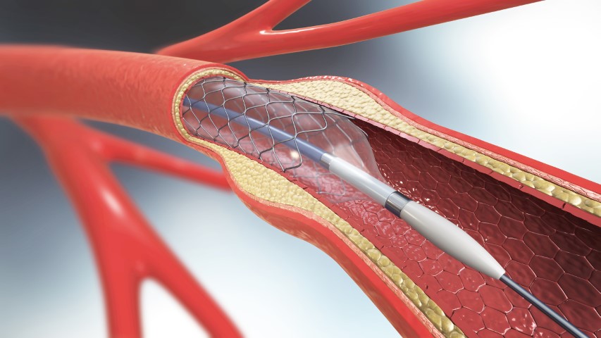
How Balloon Sinus Surgery Works

The Balloon Sinus Surgery can be performed in the ENT’s office or OT. A local or general anesthetic is applied to numb the area. Then a very small balloon catheter is inserted up through the nasal passage into the sinus. The balloon is inflated and this opens the sinus passage. The ENT flushes out the mucous and pus that has collected in the sinus with a saline solution. The balloon is removed and the sinus passage stays open.

When the balloon is inflated, it permanently alters the structure of the sinus opening. The passage not only is opened by the balloon, but it also stays open afterward. This keeps the sinus from being blocked. Because the sinus isn’t blocked it drains properly. Since the sinuses are draining you don’t get sinus headaches. It’s a cure for sinus headaches that lasts.

Balloon sinuplasty is a new technology for treating Sinus Headaches / Rhinogenic Headaches & chronic sinusitis. This is a UFDA approved technology. Affordable and inexpensive Balloon Sinuplasty procedures are available at several medical tourist destinations abroad. India, the USA, Mexico, Costa Rica, Jordan, and Turkey are using Balloon sinuplasty

Benefits of a balloon sinuplasty

A Balloon sinuplasty can treat  Sinus Headache, chronic sinusitis, opening your blocked sinuses & you will get relief. This is perhaps the biggest benefit of a balloon sinuplasty. Here are some more benefits.

1. This is a minimally invasive procedure

 The balloon sinuplasty doesn’t need an incision. The balloon is inflated and this opens the sinus passage, the cavity is cleaned and drained. No incisions or stitches!

2. The procedure takes very little time

   The balloon sinuplasty can be performed in the ENT’s office or OT, and it takes about an hour.

3. Faster Recovery

Minimally invasive procedures provide faster recovery compared to traditional surgery ― and balloon sinuplasty is no exception. The majority of patients return to their normal activities within a couple of days.

4. No nasal packing

Some nasal surgeries require nasal packing to absorb any bleeding. Remember, there’s no need for incisions or bone/tissue removal with a balloon sinuplasty, so you will not experience the same post-surgical discomfort.

5. Long-lasting relief

The most rewarding benefits of a balloon sinuplasty are that the relief is Long-lasting. In a research article published in the journal Otolaryngology-Head and Neck Surgery, patients report relief for up to two years after the procedure.

Talk To The ENT About Your Sinus Headaches / Rhinogenic Headaches

If you have recurrent sinus headaches, it’s time to talk to the ENT. Make an appointment to discuss and find out if

Balloon Sinus Surgery is the best option for you. You have nothing to lose but the pain and discomfort of sinus headaches.

What is Sinusitis?

Sinusitis/Rhinosinusitis is an infection or inflammation of the tissues lining the sinuses. It leads to poor or blocked sinus drainage. Sinusitis can occur in people of any age. Sinusitis in children also often & cause discomfort to the patient.

What are the symptoms of Sinusitis?

According to the American Academy of Otolaryngology-Head and Neck Surgery, the signs of sinusitis include:

  • Nasal inflammation
  • Nasal obstruction or Congestion
  • Pain, tenderness, and swelling around the eyes, cheeks, forehead, nose
  • Continuous Headaches
  • Runny nose
  • Thick & discolored discharge from the nose
  • Allergies & nasal polyps lead to sinus pain and symptoms
  • Loss of smell and taste, and infections

Worldwide sinusitis affects millions of people every year.

Acute vs. Chronic

Acute sinusitis lasts for a short period, as stated by the American Academy of Otolaryngology. It’s less than four weeks. During an Acute sinus infection, the patient suffers from a cold attack or other respiratory illness.

Chronic sinus infections last for more than twelve weeks. It may recur periodically. Allergies & nasal polyps are commonly noticed in these patients.

Sinusitis in Children

Sinusitis in children is a concern, and parents need to consider it seriously  & check-up with their doctor. The child’s nose needs to be examined to determine the cause of sinusitis. A mucus sample of the child may show what germ is causing the infection. It can help the doctor to opt for the appropriate treatment. Even if the child’s symptoms go away on their own, the recurring sinus attack history should be reported to the doctor’s attention. Immune problems or antibody deficiencies may be risky for chronic sinusitis.

What causes sinusitis in a child?

When the sinuses are blocked due to the infection, bacteria may start to grow. This leads to sinusitis or Nasal Congestion

The most common bacteria that cause acute sinusitis include:

  • Moraxella catarrhalis
  • Streptococcus pneumonia
  • Haemophilus influenzae

How is sinusitis diagnosed in a child?

The Doctor will record the Medical health history of the child and ask for a physical exam. The child may have tests, such as:

  • Sinus X-rays: An X-ray exam of the sinuses may help with the diagnosis.
  • CT scan of the sinuses: A CT scan shows detailed images of the sinuses
  • Cultures from the sinuses: A swab of discharge from the nose may be taken. The sample is checked for the culture of bacteria or other germs.

What are the treatments available for Sinusitis?

The ENT specialist will be the best option to treat sinusitis in a child with pain relievers, antibiotics, allergy medications, decongestants, or steroids.

According to Ann Otol Rhinol Laryngol 2009 March, Balloon Sinus Dilation Procedure in children seems to be very encouraging. There is no bone or tissue removal, the procedure seems to be suitable for use in children.

Safety and feasibility of Balloon Sinus Dilation Procedure for treatment of chronic rhinosinusitis in children

Balloon Sinus Dilation Procedure can treat chronic rhinosinusitis (CRS) by opening your blocked sinuses & you will get relief. This technique allows for restoring ventilation to the sinuses with minimal trauma to the tissues. According to Am J Rhinol Allergy Jan-Feb 2010, Balloon Sinus Dilation Procedure of the sinus Ostia in children was safe and a significant number showed improvement.

Here are some benefits of the Balloon Sinus Dilation Procedure:

1. This is a minimally invasive procedure

The Balloon Sinus Dilation Procedure doesn’t need an incision. The balloon is inflated to open the sinus passage, the cavity is cleaned and drained. No incisions or stitches are necessary.

2. The procedure takes very little time

The  Balloon Sinus Dilation Procedure can be performed in the ENT’s office or OT, and it takes about an hour.

3. Faster Recovery

Minimally invasive procedures provide faster recovery compared to traditional surgery ― and Balloon Sinus Dilation Procedure is no exception. The majority of patients return to their normal activities within a couple of days.

4. No nasal packing

Traditional nasal surgeries require nasal packing for absorbing the bleeding. Remember, there’s no need for incisions or bone/tissue removal with Balloon Sinus Dilation Procedure, so you will not experience the same post-surgical discomfort.

5. Long-lasting relief

The most rewarding benefit of a Balloon Sinus Dilation Procedure is, the relief is Long-lasting. In a research article published in the journal Otolaryngology-Head and Neck Surgery, patients report relief for up to two years after the procedure.

Talk To The ENT

If your child has chronic rhinosinusitis (CRS), it’s time to talk to the ENT. Make an appointment to discuss and find out if a Balloon Sinus Dilation Procedure is the best option for your child. Your child will recover soon if he or she gets the proper treatment from an ENT.

Heart disease often involves a problem with the valve, coronary arteries, or muscles all of which causes the heart to not function normally. The common heart disease among patients is CAD which arises from a build-up of plaque on the inner surface of the arteries. In the worst cases, it blocks the artery. Stents are instered to open the blocked artery and restor the blood by increasing the lumen area of the artery which got compromised due to plaque formation,it becomes even more important in the emergency like situation when patients suffer from the Heart Attack as due to thrombus a blood flow is totally stopped or compromised leading to the death of cardiac muscles due to lack of oxygen this need a immediate action to remove the thrombus or clot with help of medicines or angioplasty.

The idea of cardiovascular stents revolutionized the treatment of coronary-related diseases. Since then, stents have been used in coronary and peripheral procedures for decades. Let’s take a look at newer kinds of stents, their evolution and understand why better stents are needed.­­­­­­­­

What is the function of stents? 

The main purpose is to open the blocked coronary arteries which occurred due to plaque formation. A stent is a tiny mesh tube inserted into a blocked passageway to keep it open, it restores the flow of blood. It is usually needed when plaque blocks blood vessels. Commonly used heart stents  are made up of materials like stainless steel,cobalt chromium or platinium. It can be coated with medication to facilitae optimal healing.

Evolution of early stents

The first used stents were successful at reducing the risk of restenosis following angioplasty by about half, from 20% to 10% in the 12 months following the procedure. The first generation of stents was called bare-metal stents (BMS) as they were mostly fabricated from metal. These permanent frameworks are made from chrome steel and cobalt-chromium alloys for balloon-expandable and Nickel-titanium for self-expanding stents. This revolution was considered the turning point in the field of angioplasty however it had its disadvantages of increased risk of restenosis.

Stent developers started to coat bare-metal stents with polymers containing drugs that were aimed at inhabiting extra tissue growth at the site of the stent to reduce the rate of restenosis. These stents are called drug-eluting stents (DES). DES has been constantly developed by using several drugs.

DES has bought its advantage to overcome the limitation of BMS. Despite the influence in the clinical practice in vascular intervention, the major subject of the debate was incomplete endothelialization and hypersensitivity reactions to the polymer coating. The first-generation DES had all these drawbacks which had them failed. Due there coating material not being biodegradable or biocompatible their long-term efficiency was questioned. The potential risk associated with DES were late thrombosis and delayed healing.

The formation of a grume within the artery at the location of the stent is stent thrombosis. It is different from restenosis. Restenosis is the regrowth of tissue, it is a problem but it grows gradually so when it occurs there is time to treat it. In contrast, stent thrombosis tends to occur suddenly without warning, it commonly leads to the complete occlusion of the artery that then tends to produce a myocardial infarction (Heart attack) or sudden death.

In the youth of stenting, the matter of early thrombosis was recognized and successfully addressed by administering powerful antiplatelet or blood thiner for a couple of months after stenting. But within few years of the widespread use of DES the problem of late thrombosis was discovered. Late thrombosis may be a sudden thrombosis within the site of the stent that happens a year or two after the procedure. It is as catastrophic as early stent thrombosis. Hence the cardiologists started prescribing antiplatelet therapy for up to a year or for longer period of time. The strong use of anti-platelet drugs carried the risk. The problem of late stent thrombosis led stent developers to a challenge to develop a new type of stent that reduces this problem or eliminates it.

Researchers now believe that the polymer coating on DES may themselves increase inflammation and delay healing. Now several companies are identifying the problem and trying a general approach keeping this in mind.

Advanced stent technology  

Better durable polymers DES appear to cause less inflammation and also allow better healing at the site of treatment. These stents are referred to as second-generation DES, the newer stent design use cobalt-chromium which has greater radial strength per thickness and is radio-opaque, and thus allows thinner struts.

Bioabsorbable polymers

DES has been available for several years, they employ a polymer coating which is absorbed within few months leaving just the bare stent. These stents offer the benefits of DES for several months then they become BMS. This reduces the risk of late thrombosis. As compared to the first generation DES the risk of late thrombosis is reduced with both second-generation DES and bioabsorbable polymers DES. But there is no indication that bioabsorbable polymer performs better than the second-generation DES. They both still require very prolonged therapy and anti-platelet tests.

Bioresorbable stents

Bioresorbable stents also called as bioresorbable scaffold, biodegradable stent, or naturally- dissolving stent they are made from a material that may dissolve or be absorbed in the body. The main purpose of the bioresorbable stents is to overcome the drawbacks of DES and BMS. Currently, A metallic drug-eluting stent remains in the body as a permanent implant which can lead to an increased risk of complications year on year. It may pose a hindrance if the future procedure needs to be performed in the same artery. But on the other hand, BRS provides a temporary scaffold to the lesion to restore the blood flow, once the blockage is treated and healed completely BRS dissolves on its own leaving the artery in its natural state with no foreign residue. Hence providing both physicians and patients the room to explore further treatment, if required in the same blood vessel.    

We get to see all these extraordinary engineering in stent technology, it seems like sooner or later we might develop stents that would be able to eliminate the risk of both restenosis and thrombosis. But you need to make sure that before you agree to a stent talk to your doctor and explore options whether it will be helpful for you.

The possibility of having heart disease or a heart valve condition is overwhelming and frightening for most people. That is why having an understanding of heart diseases is important so that one can be well equipped with the right information, and hence take the right steps towards treatment.

Heart valve conditions are usually due to birth defects or age-related issues. Many heart valve conditions can be first identified with the help of a murmur, that is a whoosing sound as blood flows from one chamber to the next, or it may sound like an extra click when a valve allows backflow.

A murmur may indicate conditions such as Aortic stenosis, prolapse, regurgitation, etc. While medicines can be prescribed for a heart valve condition, they are mainly for the following reasons

  • To reduce unpleasant symptoms from milder forms of the disorder.
  • To maintain heart rhythm if a related arrhythmia is present.
  • To lower the patient’s risk for clotting and stroke.

Valve diseases are generally progressive by nature, and the outlook for those who do not receive adequate and timely treatment could be fatal. If you are suffering from an aortic valve disease it is highly advisable to opt for a surgical procedure for low surgical risk patients.

The heart has four chambers. The two upper chambers are called the left and right atrium and the lower chambers are called the left and right ventricle. The four valves that are at the exit of each chamber carry out the function of continuous blood flow through the heart to the lungs and the rest of the body. The four valves are the tricuspid valve, pulmonary valve, mitral valve, and aortic valve.

The aortic valve controls blood flow from the left ventricle into the aorta (the main artery in your body). When this valve opens, the oxygen-rich blood is pumped to the aorta and then to the rest of the body. It is key to the body’s blood circulation system.

Reasons you might need to undergo heart valve surgery

Types of aortic valve disease that may require treatment with aortic valve repair or replacement include the following:

Congenital heart disease: This condition may contribute to other complications that prevent the aortic valve from operating properly. For instance, a person may be born with a condition where the aortic valve does not have enough tissue flaps (cusps) or with a valve that is the wrong size or shape, or without an opening that allows blood to flow normally.

Aortic valve stenosis: When the aortic valve becomes narrow or obstructed, it becomes difficult for the heart to pump blood into the aorta. This may be caused by the thickening of the valve’s closure flaps, congenital heart disease or post-inflammatory changes associated with rheumatic heart disease. Around 75% of patients with unoperated aortic stenosis may die 3 years after the onset of symptoms.

Bicuspid aortic valve: A birth defect where only two cusps grow instead of the normal three. This is a common cause of aortic stenosis. Another cause may be that the valve opening does not grow as the heart does. This makes it harder for the heart to pump blood to the restricted opening. The defective valve becomes narrow and stiff over time because of a calcium build-up. Age-related Aortic Stenosis usually begins after age 60, but often doesn’t show symptoms until ages 70 or 80.

Aortic valve regurgitation: When the blood flows backward through the aortic valve into the left ventricle each time the ventricle relaxes rather than in the normal one-way direction from the ventricle to the aorta. This may be caused due to an abnormal valve shape present at birth (congenital heart disease) or by a bacterial infection.

Most symptomatic patients of AR will require valve replacement surgery within 2 to 3 years of developing breathlessness and symptoms of heart failure. A Swedish study has proven that these individuals have a life expectancy of 2 years lesser than the healthy population of the same age.

Valve diseases, although gradual and progressive are fatal if not treated in the right manner. Valve replacement surgery is recommended as it helps alleviate your symptoms and add quality years to your life.

Careful monitoring and doctor supervision may be all that is needed for a few people with mild aortic valve disease without symptoms. However, in most cases, aortic valve disease and dysfunction can get worse.

Such conditions eventually require surgery to reduce the risk of complications such as heart failure, heart attack, or a stroke or death by cardiac arrest. This is when a doctor will recommend heart surgery.

What are the consequences of not having a heart valve surgery?

  • Decreased life expectancy and quality of life

As per an estimate, as the aortic valve disease gradually worsens, the average rate of survival without undergoing surgery is a mere 50% post 2 years, while it is 20% post 5 years.

While you may feel normal and not notice any problems for years, valve disease is not a condition to be ignored. Once you begin to experience symptoms like the ones mentioned above, not only does life expectancy and quality of life decline but the severity of diseases such as aortic heart stenosis increases.

  • Contributes to more health complications

As the wall of the left ventricle works harder to pump blood through the narrow valve

opening into the aorta, the wall might show muscular thickening, along with the symptoms of aortic stenosis like fatigue and weakness.

The thickened wall allows less room for an adequate amount of blood circulated to the body, as it takes up more space inside the lower heart chamber. This may cause heart failure.

It is advisable to undergo valve replacement surgery as it not only adds to a better quality of life and solves the issue of aortic stenosis but also helps avoid the consequential conditions that come along with it.

Coping with Aortic Stenosis can not only be stressful physically but also mentally. Learning about your condition and the right treatment by talking to your doctor about it can help a great deal. Aortic Stenosis can be fatal and not undergoing the right treatment or prescribed surgery can lead to not just a deterioration in your condition but also create newer complicated conditions for your health. Knowing and understanding your condition as well as the benefits and success rates of the treatment gives you an upper hand and a better chance at survival.

Disclaimer: This blog is only for awareness purposes. We do not intent to promote any medications given in the blog. Please consult your physician before taking any medication.