Category

Surgery:

Category

The news that one might be suffering from a heart condition that requires a heart stent can be stressful to process. It is important to understand the reasons why your doctor might have recommended a stent in the first place.

Arteries are blood vessels that carry out the function of circulating blood away from the heart to the rest of the body. They are shaped like tubes and the pumping contractions of the heart propel the blood through the arteries.

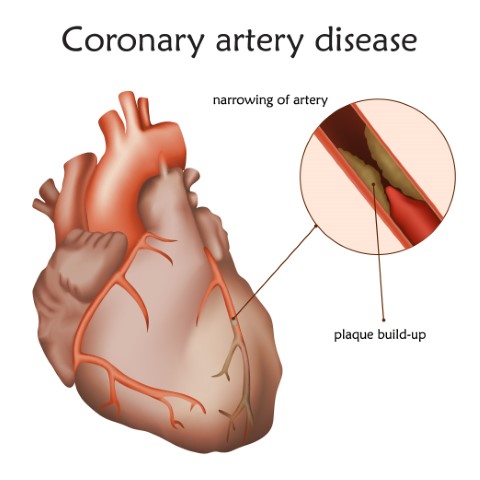
When a buildup of plaque i.e cholesterol, fat, and other substances collect in the bloodstream, it sticks to the walls of the arteries, and over time, this buildup narrows the arteries, limiting the amount of fresh blood that can be circulated to the body. 

Over time, people with narrowed coronary arteries may begin to notice symptoms such as chest pain. This buildup of plaque in the coronary arteries is a cause of coronary heart disease. People with this condition may be at a higher risk of many serious complications such as a heart attack or a stroke if they do not receive treatment.

What is Coronary Artery Disease? Coronary Artery Disease (CAD) is the most common form of heart disease. It occurs when one or more of the coronary arteries becomes narrow, thus restricting the flow of blood to the heart muscle. Without adequate blood, supply the heart becomes starved of oxygen and vital nutrients required for the proper functioning of the heart muscle which may lead to chest pain called angina. The process is often called hardening of the arteries or atherosclerosis.

Symptoms of CAD may include

Early warning signs such as

  • A squeezing, suffocating, or burning feeling in your chest tends to start in the center of your chest but may move to your arm, neck, back, throat or jaw.
  • fatigue
  • pain
  • dizziness

Symptoms in women are comparatively non-traditional:

  • vague chest discomfort
  • fatigue
  • indigestion
  • Anxiety
  • sleep difficulties

If left untreated, CAD can lead to other serious problems such as heart attack, stroke, or even death.

When a coronary artery is narrowed by a buildup of fatty deposits called plaque, it can reduce blood flow, causing chest pain. If a clot forms and completely blocks the blood flow to part of the heart muscle, it can result in a heart attack.

Therefore if the artery is at risk of collapsing or becoming blocked, doctors may recommend inserting a stent to keep it open.

What is a stent?

A stent is a tiny wire mesh tube that your doctor can insert into a blocked artery passageway to keep it open. Its function is to open the blocked artery and post-treatments stents remain permanently in the body. Stents are made of either metal or plastic. Stents can also be coated with medication to help keep a blocked artery from closing.

How are stents used?

The stent is mounted on a balloon catheter. It is tracked through the artery to the narrowed segment/blockage which is then moved into the area of the blockage. The balloon is then inflated resulting in the stent expanding and locking in place, forming a scaffold that holds the artery open. The balloon is then deflated and withdrawn.

The stent stays permanently and improves blood flow to the heart muscle. Stents are used based on the features of the artery blockage such as the size of the artery and where the blockage is.

What are the advantages of using a stent?

  • After balloon angioplasty or other procedures that use catheters, certain patients may experience a re-narrowing of the artery. Stents help to reduce the re-narrowing.
  • Patients who have angioplasty and stents have less discomfort and also recover faster from these procedures as compared to patients that choose to undergo coronary artery bypass surgery (CABG)

How long will a stent last? Are stents permanent?

Commonly used metallic stents are permanent and stay in the body for lifelong.

Re-narrowing (Restenosis) of the treated coronary artery can occur in some cases and may require a re-intervention in the future. However, with the latest generation, the drug-eluting stents’ restenosis rate has reduced significantly.

Bioresorbable scaffold or stent is the latest innovation in the field of stent technology. Unlike metallic stents, they don’t stay permanently inside the patient body. Post healing, they dissolve naturally in 2-3 years thus leaving the artery in its natural state.

What happens after stent insertion?

Depending on the insertion site, there may be soreness, for which mil painkillers are usually given. Your doctor might prescribe medication to prevent clotting, and might want you to remain at the hospital overnight. This helps ensure that there are no complications. In cases of coronary events like a heart attack or a stroke, you might be asked to stay for a longer time. Depending on the severity of the cause of the stent insertion, such as a heart attack, recovery might take a while. Until then it is advised to take it easy for a couple of weeks, avoid heavy lifting, etc.

Are there any risks involved in the insertion of stents?

Like any other medical procedure, Stenting may also involve risk, but with the latest advancement in technology and the treatment procedure, it has been minimized significantly. One should discuss and understand with their Physician about the common risk factors which is involved with stenting.

Some of the common risk factors may include bleeding from the treatment site like groin from where the catheter was introduced, allergic reaction to dye or stent, re-narrowing of the artery or clot formation, etc

Insertion of a foreign body in your blood, there is a one in 50 chance of blood clots forming on the stent. Your physician may prescribe certain medications to lower the risk of blood clots. 

While a stent can result in remarkable improvement of your condition, it is not a cure for heart disease. You will need to address the contributing factors like high blood pressure, cholesterol, and being overweight as well. Eating a well-balanced diet, exercising regularly, even quitting smoking can lead to a heart-healthy lifestyle.

Disclaimer: This blog is only for awareness purposes. We do not intent to promote any medications given in the blog. Please consult your physician before taking any medication.

Hip replacement is a surgical procedure in which a surgeon removes the damaged and painful sections of the hip joint and replaces it with an artificial one that is usually made from metal, ceramic, and High density  plastic components. This procedure is undertaken to reduce pain and improve one’s joint movement and function. The artificial joint is known as a prosthesis.

Hip replacement is usually done when other, less invasive methods have failed to provide relief to the patient. Also known as Hip Arthroplasty, hip replacement is also considered if the hip pain interferes with one’s daily activities. Damage caused due to Arthritis is one of the most common reasons for Hip Replacement Surgery. Some conditions that may require hip replacement surgery to treat it are:

Osteoarthritis: Also known as wear-and-tear arthritis, this condition damages the protective cartilage that cushions the ends of your bones which helps the joints move smoothly.

Rheumatoid Arthritis: This is an autoimmune disease in which there is inflammation in the joints, which in turn leads to bone erosion and joint deformity.

Osteonecrosis: Osteonecrosis occurs when there is no adequate blood supply to the ball portion of the hip joint, which might occur due to dislocation of the hip bone or hip fracture.

Does an artificial hip work better after a hip replacement?

Hip replacement offers the greatest benefit of pain relief. Along with that, there is an improvement in movement, strength, and coordination of the lower body. It also helps you to get back to your active life. But, despite being highly effective, an artificial hip is associated with certain limitations.

1. Durability: Though majority the of the artificial hip implants last for 15 to 20 years, one cannot guarantee smooth functioning in every case of certain situations such as infections or trauma, an artificial hip can get worn out quickly, making it necessary for a second replacement. Normally, an artificial hip can work for up to 15 – 20 years, without any major trouble.

2. Susceptibility to allergy: Though these problems are rarely seen, they can occur. The patient may display signs of metal allergy after the surgery. This is characterized by skin rash, itching sensation, and discoloration in the area where the artificial joint has been fixed. 

3. Weather problems: In some patients, the natural bones surrounding the hip implant take more time to adapt to the metallic nature of the hip, leading to increased weather sensitivity in the patient. Due to this phenomenon, a patient may feel increased pain and stiffness in the artificial hip in certain seasons like monsoon or winter.

4. Caution in future dental visits: You need to take care while visiting your dentist after your hip replacement as you could catch an infection. Preferably, you should take a prophylactic antibiotic.

What type of permanent restrictions will one have after a hip replacement surgery?

Every patient’s experience with Hip Replacement Surgery is not the same. Hence, it is not possible to predict whether a particular patient will have permanent movement restrictions after hip replacement or not. Most of the patients can return to a normal daily routine without any permanent restrictions, albeit with less pain and discomfort. However, a risk always prevails regarding an implant getting worn out, in which case the patient might need a second replacement. Though there are no permanent restrictions in most cases, there are certain activities one has to avoid for a certain period. These restrictions are also known as posterior hip precautions. They are:

  1. Cross your legs: One should not cross their legs past the midline of the body after a hip replacement surgery. To avoid crossing your legs, the surgeon may also advise sleeping on your side or keeping a pillow between the legs.
  2. Forward bending: It is also important to ensure that you do not bend your hip beyond 90 degrees after a hip replacement. Your knee must be below your hip joint. You can utilize cushions and pillows to elevate your seat, to ensure that your knees are positioned lower than your hips.
  3. Positioning your feet: Ensure that you keep your feet and your knees pointed straight ahead, in the front. Do not try turning them in or out. Also, avoid sitting in the same position for more than 30 40 minutes.
  4. Seating arrangement: Make sure that you are using a firm chair with a straight back and armrests. Avoid chairs that are too soft, rocking chairs, recliners, and stools.
  5. Climbing the stairs: Avoid climbing the stairs very frequently, as it will put a strain on the artificial joint.

Even after the joint has completely healed strenuous physical activities like sports must be avoided. It is important to keep in mind that the prosthetic joint has only been designed to withstand day-to-day physical activities. You can work with a physical therapist to learn various techniques and guidelines while performing various activities. If you do not follow the above-mentioned restrictions, you may run the risk of dislocating your joint, leading to second replacement surgery. Hence, you must be well-informed and take precautions with utmost care as suggested by your surgeon.

The human heart has four chambers, with the upper chambers being the left and right atrium and the lower chambers being the left and right ventricles. The heart valves are present at the end of each chamber and they maintain the one-way blood flow from the heart to the lungs and the rest of the body. The four different valves are – Pulmonary Valve, Tricuspid Valve, Aortic Valve and Mitral Valve.

The tricuspid valve is placed between the right atrium and right ventricle. Through this valve, blood flows to the right ventricle. The function of the pulmonary valve is to control the flow of blood between the right ventricle and the lungs. The mitral valve is positioned between the left atrium and left ventricle and allows the blood to flow between them. The aortic valve controls the blood flow from the left ventricle to the aorta. The aorta is the main artery of the body, from which blood flows to the other parts of the body.

What are Valvular Diseases?

Valvular diseases, commonly known as heart valve diseases, occur when one or more of the heart valves do not open or close properly, affecting the blood flow. When more than one valve is affected, it is known as Multiple Valvular Heart Disease. The different types of valvular diseases are:

Stenosis:

In this condition, the heart valves stiffen, leading to the narrowing of the valves. This in turn restricts the blood flow from the valves to the other organs. It keeps the valve from opening fully to allow smooth blood flow, due to which an inadequate amount of blood is supplied to the body. In severe cases, the valve opening may completely close, due to which the blood flow is blocked.

  • Tricuspid valve stenosis: In this condition, the blood is unable to flow from the right atrium to the right ventricle, causing the atrium to enlarge. This affects the blood pressure and flows in the surrounding chambers. It can also cause the right ventricle to shrink or become smaller, restricting the blood circulation to the lungs.
  • Pulmonary valve stenosis: If the pulmonary valve narrows, the blood flow from the right ventricle to the lungs is restricted, due to which oxygen-rich blood cannot be pumped to the rest of the body. In this condition, the right ventricle might have to compensate by pumping blood harder through the narrowed valve, increasing the pressure on the heart.
  • Mitral valve stenosis: In this condition, the blood flow from the left atrium to the left ventricle is reduced, causing fatigue and shortness of breath. It may also cause enlargement of the atrium and fluid build-up in the lungs.
  • Aortic valve stenosis: This is one of the most common heart conditions. In this condition, the blood flow from the heart to the aorta and the rest of the body is restricted. Due to this, the left ventricle has to pump harder to ensure blood flow to the aortic valve. This may also lead to the thickening of the left ventricle.

Prolapse:

Valvular prolapse is a condition in which the valve flaps slip out of place or bulge. This can lead to the valves being unable to close properly. Due to this, blood may flow backwards through the half-closed valve, disrupting the one-way blood flow.

  • Mitral valve prolapse: Also known as Barlow’s syndrome, click-murmur syndrome and floppy-valve syndrome, this condition prevents the valve from closing evenly. The mitral valves may also bulge upwards into the atrium when the two ventricles contracts. Due to this, a small amount of blood may flow backwards through the valve.
  • Tricuspid, Pulmonary and Aortic valve prolapse: These prolapses are less common as compared to Mitral valve prolapse. In the above-mentioned conditions, the leaflets/flaps of the valves do not close properly to form a tight seal.

Regurgitation:

Regurgitation happens when the valve doesn’t close properly, allowing the blood to flow backwards. This disrupts the one-way blood flow and puts a strain on the heart. This reduces the efficiency of the heart in pumping oxygen-rich blood to the rest of the body.

  • Tricuspid valve regurgitation: In this condition, the blood pumped from the right ventricle to the lungs may flow back to the right atrium, leading to the enlargement of the atrium.
  • Pulmonary valve regurgitation: In pulmonary valve regurgitation, the pulmonary valve doesn’t close properly, leading to the blood flowing backwards from the lungs to the heart. This backward blood flow may cause oxygen-rich blood to blend with oxygen-poor blood, reducing the availability of oxygen-rich blood to the rest of the body.
  • Mitral valve regurgitation: Mitral valve regurgitation can be a result of mitral valve prolapse. In this condition, blood leaks backwards into the left atrium through the mitral valve. In severe cases, this condition may lead to the enlargement of the atrium and lung congestion.
  • Aortic valve regurgitation: In this condition, blood leaks backwards from the aorta to the left ventricle, due to which the body does not get an adequate amount of blood, leading to the heart pumping harder to compensate. This may cause the thickening of the left ventricle.

Symptoms of Valvular Heart Diseases:

Some common symptoms of heart valve diseases are:

  • Discomfort or tightness in the chest.
  • Palpitations are a common symptom of heart valve diseases, which may cause the heart to enlarge, leading to irregular heartbeats (arrhythmia).
  • Shortness of breath – as valvular diseases disrupt the flow of oxygen-rich blood to the body, causing breathlessness.
  • Fatigue and weakness
  • Dizziness and fainting
  • Swelling occurs when the blood flows backwards, leading to fluid buildup and congestion, which in turn leads to swollen abdomen and feet.

Though the symptoms may slightly vary for different valvular diseases, it is important to be mindful of them and connect with your doctor even if you experience the mildest of symptoms. Though severe conditions may require surgical procedures, a few lifestyle changes will help keep these heart valve conditions at bay. Be aware and take precautions!

What are Piles/Hemorrhoids?

Piles, also known as Hemorrhoids, are a collection of swollen tissues and veins in your anus and lower rectum. They can be of two types – internal and external. Internal hemorrhoids develop inside the anal canal, while external hemorrhoids develop on the skin around the anus. These hemorrhoids may vary in size. Piles may occur due to constipation, chronic diarrhea, lifting heavy weights, pregnancy, and straining while passing stools.  

Usually, hemorrhoids don’t require any treatment and resolve on their own. But when the condition turns severe, treatment is needed. The symptoms of piles depend on the type and location. In the case of external hemorrhoids, the symptoms might include irritation/itching in the anal region, pain, discomfort, swelling, and bleeding. In the case of internal hemorrhoids, the hemorrhoids cannot be seen, but they can cause painless bleeding while passing stools. Protruding hemorrhoids have feeling of some mass in the anal region that causes pain and irritation.

What are the various treatment/cure methods for Piles?

There are different ways to cure piles, depending on the severity of it. Some of the most effective treatment methods are:

For early and mild pile piles:

Home Remedies: If the hemorrhoids are mild, then you can cure them through certain home remedies such as making dietary and lifestyle changes, using over-the-counter hemorrhoid cream containing hydrocortisone, and soaking your anal area in warm water for 10-15 minutes every day.

Rubber Band Ligation: In this procedure, your doctor will place one or two tiny rubber bands on the base of the internal hemorrhoids to stop the circulation. Within a week, the hemorrhoid withers and falls off.

Sclerotherapy: Your doctor will inject a chemical into hemorrhoid to shrink it. Though this procedure causes less pain, it is also less effective than the rubber band ligation.

Coagulation: This technique uses laser or infrared light to harden internal hemorrhoids that bleed, shriveling them.

For moderate to severe piles:

External Hemorrhoid Thrombectomy: In case of thrombosis (a painful blood clot) in external hemorrhoid, your doctor will remove hemorrhoid, which will provide immediate relief. This procedure is done under local anesthesia and is extremely effective if performed within 72 hours of the blood clot.

Hemorrhoid Removal: Also known as Hemorrhoidectomy, this is a surgical procedure in which the surgeon removes the excess tissue which is protruding out of anal canal that causes bleeding. This method is used to treat severe piles.

Hemorrhoid Stapling: This procedure is also known as Stapled hemorrhoidopexy. It blocks the blood flow to the hemorrhoid tissue by taking cutting and pulling the piles up on its position. It is usually used to treat internal hemorrhoids.

What is Anal Fissure?

An anal fissure is a tear in the mucosa (a thin, moist tissue) lining of the anus. The tearing of the skin causes severe pain and bleeding during and even after bowel movements. Though it is most commonly seen in infants and children, an anal fissure can affect people of all age groups. Most times, the fissure heals on its own with a month to six weeks. But if it persists after that, it can be considered chronic. Sometimes, the fissure tends to be deep enough to expose the muscle tissue.

The most common causes of an anal fissure are constipation, straining during bowel movements, passing large/hard stools, diarrhea, childbirth, and anal intercourse. A person suffering from an anal fissure may experience one or more of the following symptoms, which include, mild/severe pain during bowel movements, visible tear/crack on the skin around the anus, a small lump near the tear, streaks of blood after passing stools on the toilet seat or the tissue paper, pain after bowel movements that lasts for a long time.

How can Anal Fissure be cured?

Usually, Anal Fissure doesn’t require extensive treatment. Certain home remedies can help cure anal fissure and relieve symptoms. Some treatment methods to cure anal fissure are:

Home remedies: You can take measures to keep your stools soft, such as increasing the intake of fibers and fluids, and taking a sitz bath to relax the anal muscles, relieve irritation, and increase blood flow.

Applying Nitroglycerin: Applying a nitroglycerin cream or ointment will help increase blood flow to the anal area and reduce inflammation.

Botox Injection: The Botulinum toxin type A (botox) injection may help paralyze the anal sphincter muscle and relax the spasms.

Blood Pressure Medicines: These medicines can also help relax the anal muscle. This can be used when nitroglycerin is not effective or causes significant side effects.

Surgery: If the anal fissure is severe, your doctor might suggest surgery known as the Lateral Internal Sphincterotomy (LIS), in which a small portion of the anal sphincter muscle will be removed to reduce pain and promote healing.

What is the difference between Piles and Fissure?

While piles are a collection of swollen tissues in and around the anus region, fissure refers to a crack in the anal cavity.

What is Anal Fistula?

An anal fistula is a small tunnel that develops between the anal canal and the skin near the anus. They’re usually the result of an infection near the anus causing a collection of pus (abscess) in the nearby tissue. When the pus drains away, it can leave a small channel behind. In the fistula, the center portion of the anus gets infected and forms an abscess, which starts oozing pus.

What is the difference between Piles and Fistula?

Piles are caused due to constipation, diarrhea, pregnancy, and weight lifting, while fistula is caused due to trauma, Crohn’s disease, warts or cancer. Piles can usually be diagnosed by self by observing the signs and symptoms while detecting fistula might be difficult and may require an MRI scan or a sonofistulagram.
Piles are less severe as they can be cured with home remedies and other less invasive techniques, while fistula can only be treated through surgeries such as fistulotomy, seton techniques, advancement flap procedure, and LIFT procedure. Video-Assisted Anal Fistula Treatment (VAAFT) is a non-surgical method commonly used to treat fistula.

Be it piles, anal fissures or fistula, the first step to prevent them is by practicing better hygiene, especially when it comes to defecation. All three conditions can also be prevented by indulging in a high-fiber diet and increasing the intake of fluids, everyday physical activities and avoiding strains. It is also important for one to be mindful of the symptoms and seek immediate medical help to prevent the conditions from becoming severe.

Disclaimer: This blog is only for awareness purpose. We do not intent to promote any medications given in the blog. Please consult your physician before taking any medication.

Oral Cancer, as the name itself suggests, refers to cancerous growth in any part of the mouth (oral cavity). It falls under a category of cancers called Head and Neck Cancers. Also known as Mouth Cancer or Oral Cavity Cancer, this type of cancer occurs on the lips, gums, hard and soft palates, inner cheek lining, tongue, the roof of the mouth, under the tongue, and throat. Mouth Cancer can be life-threatening if not diagnosed and treated immediately. Getting quarterly or biannual dental checkups can help in the early detection and treatment of oral cancer.

Make an appointment with your doctor as soon as you spot symptoms such as a lip/mouth sore, a white or red spot in your mouth, a lump in the mouth, difficulty/pain while swallowing, or pain in the mouth. The doctor will ensure if the symptoms are due to other causes such as an infection or any other underlying condition before diagnosing oral cancer.

Who are at risk of getting diagnosed with Oral Cancer?

According to the American Cancer Society, men are at a higher risk of getting diagnosed with oral cancer than women. Oral cancer is most prevalent in men over the age of 40-50. Some of the risk factors that lead to the development of oral cancer include:

Smoking: People who have the habit of frequently smoking cigarettes, cigars, or pipes are more likely to develop oral cancer as compared to people who do not smoke.

Other forms of tobacco use: Apart from smoking, people who consume tobacco by snuffing and chewing are prone to oral cancer in the gums, lips, and inner cheeks.

Alcohol consumption: People who consume excessive amounts of alcohol are six times more likely to get diagnosed with oral cancer as compared to non-drinkers.

Family History: If one or more of your family members are diagnosed with oral cancer, you are more susceptible to the disease.

Weak immune system: It is your immune system that helps your body fight against various illnesses. If your immune system is weak due to certain conditions, you are prone to oral cancer, along with other diseases.

Human Papillomavirus (HPV): The Human Papillomavirus (HPV) is a sexually transmitted virus that could be a major contributing factor for the development of oral cancer.

What are the precancerous conditions of the oral cavity?

A precancerous condition is a condition that involves abnormal cells which lead to an increased risk of developing cancer. Depending on the type of cancer, the precancerous conditions can be few or many.

In oral cancer, precancerous conditions are the changes that take place in the cells of the mouth. These conditions are not developed into cancer yet. But, if left untreated, there are higher chances that these abnormal changes or mutations might advance into oral cancer.

Oral cancer has the two most common precancerous conditions. They are:

Leukoplakia:

Leukoplakia is an unusual grey or white patch that is visible on the tongue, the gums, the inner cheek linings, or the floor of your mouth. If one has leukoplakia, it is not necessary that he/she will develop oral cancer. Depending on the size and shape of the abnormal cells, this precancerous condition may develop into oral cancer.

Erythroplakia:

Erythroplakia is an abnormal red patch or a group of red spots that is visible on the mucous membrane in the mouth, with no definitive cause. This precancerous condition has higher chances of developing into oral cancer as compared to Leukoplakia. Around 50% of these conditions advance into Squamous Cell Carcinoma.

How can you prevent oral cancer?

Though there are no proven measures to prevent oral cancer. However, there are certain precautions one can take to reduce the risk. They are as follows:

Stopping the consumption of tobacco: As mentioned above, tobacco usage is one of the most common risk factors that lead to oral cancer. If you are a regular consumer of tobacco, put a stop to it. Smoking, chewing, or snuffing tobacco is dangerous as it exposes the cells in your mouth to cancer-inducing chemicals.

Moderate alcohol consumption: Excessive alcohol consumption can weaken the cells in your mouth, making you vulnerable to oral cancer. Hence, alcohol should be consumed in moderate amounts or to be completely avoided to lower the risk of mouth cancer.

Avoid excessive sun exposure: Protect the skin on your lips from excessive sunlight by applying lip balm with sun protection as a part of your regimen. You can also wear a broad-rimmed hat when you step out and stay in the shade as much as possible.

The first step to fighting oral cancer is to be mindful of the risk factors and symptoms. Although oral cancer can become fatal if not treated on time, taking precautions to reduce the risk will help keep oral cancer at bay. Have a regular quarterly/biannual check-up with your dentist and ensure proper oral hygiene, because prevention is better than cure!