Category

Surgery:

Category

Pregnancy is an exciting time for an expecting couple, especially if it is their first child. Most couples wish to have a normal delivery as it is the safest way to have a child. But many times, a woman or the infant inside her may suffer from certain ailments that can cause complications during the pregnancy. At such times, doctors may recommend opting for surgical delivery method, which is a Caesarean delivery.

A caesarean or C-Section delivery is a surgical delivery process that is usually performed when normal vaginal delivery isn’t safe for either the mother or the child.

To start, What is a C-Section or Caesarean delivery?

When you put it across simply, a C-Section delivery, also known as Caesarean delivery is when the doctor makes a cut over the skin and on the lining of the uterus to make the delivery process a bit easier. Different types of incisions, either vertical or horizontal are done. It is a quick, often painless and effective surgical delivery method that doctors choose for women who have complicated pregnancies.

What types of C-Sections can one undergo?

During pregnancy, visits to the doctor for prenatal care need to occur frequently. In these sessions, the doctor may spot a complication for which you may need to forego the natural or normal delivery route and in its place, opt for a C-Section (Caesarean) delivery. Largely, the decision to undergo a C-Section is taken by the doctor when the actual delivery is taking place. Such intervention is called an Emergency C-Section.

But, in a few cases, the doctor may tell the expecting woman beforehand that she has to undergo a caesarean to avoid any complications that may arise if a normal delivery is attempted. Sometimes, there may not even be any complications. A woman may just want to avoid the pain that comes with labour and opt to go for a C-Section delivery instead. But of course, the decision to undertake any type of delivery surgery eventually lies with the doctor.

What C-Section is recommended for women?

Some couples may want to go for a planned C-Section, but the decision to do so lies with the doctor, who decides in the best interest of both mother and child. The few instances where a C-Section is the preferred choice include:

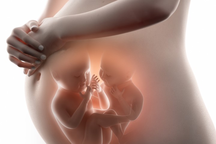
1) Instances of multiple births, where the women give birth to either twins or triplets. While doing so, the doctor may go for a caesarean as normal vaginal delivery can get quite complicated or tiring.

2) Instances where the expecting mother has an underlying chronic medical condition such as diabetes or hypertension, which can lead to serious problems, especially while delivering the child.

3) Some women may suffer from an infection or sexually transmitted disease such as HIV. In such cases, the yet to be born infant may also be afflicted and as such, a normal delivery may not be the safest option.

4) A C-section may also be needed in some situations, such as delivering a very large baby in a mother with a small pelvis, or if the baby is in a feet-first position.

5) Sometimes the doctor’s decision to perform a C-section is not planned, and it is performed for emergency reasons as the health of the mother, the baby, or both of them is at risk because of troubles during pregnancy or after a woman has gone into labour.

What happens after a C-Section Surgery?

Though C-sections are generally safe and help in overcoming a lot of complications during the delivery, there is always a possibility of complications arising from it. Unlike a natural vaginal delivery, the recovery time for caesarean delivery is much higher, both in hospital and afterwards. Certain complications can arise from a C-Section surgery. These include excessive bleeding, bowel injury or the chance of contracting an infection. Fortunately, serious complications from caesarean deliveries are rare.

Are there any effects on the infants?

While a C-Section surgery is done in the best interest of both the mother and the child, there are certain issues that the infant may face as an outcome of caesarean delivery. Some of these effects include:

1) Altered Gut Bacteria: This can create problems in the development of a healthy immune system in babies which makes them susceptible to illness.

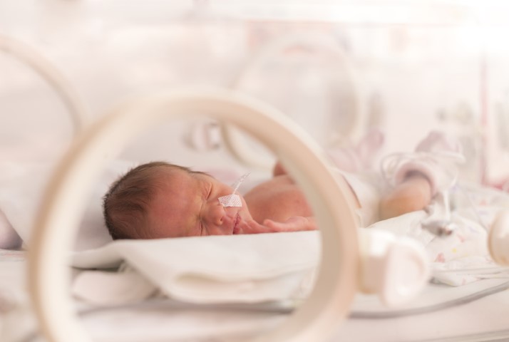
2) Delayed skin-to-skin contact:  Usually after caesarean delivery, the infant is kept in an incubator. The lack of touch causes problems in the transfer of healthy bacteria from the mother’s skin to the baby, maintenance of body temperature and delayed breastfeeding initiation. Along with these physiological effects, there is also the psychological brunt of delayed parent to baby bonding.  

3) Excessive fluid accumulation: A complication that takes place during delivery that can cause obstructions in breathing for the newborn infant.

4) Non-molding of baby’s head: If your baby is born breech (buttocks or feet first) or by caesarean delivery (C-section), the head is most often round and not moulded.

5) Formation of Organs: A caesarean delivery can occur pre-term, which is, before the normal 37 week period. Due to this, the infant’s organs may not be fully formed and lead to complications post its birth.

6) Risk of asthma, obesity and delaying of developmental milestones later in life.

In today’s time, both science and medicine has evolved exponentially. So, if your doctor does suggest you to go through a C-Section delivery, there isn’t much that you need to be worried about.

If we look purely at statistics, about one-third of the deliveries worldwide are taking place via C-Section, with quite a good chunk of them being planned C-Section surgery. Of course, natural vaginal birth is always the most preferred choice, but it is always good to look at your options if complications arise. If you are still in the planning stage, it is also advisable to get yourself checked for any pregnancy-related complications, and when a woman does get pregnant, prenatal follow-ups with a doctor is a must. While the choice of delivery, either natural or surgical is best left in the hands of your doctor, there should be no reason to worry about C-Sections, as it will enable you to have a healthy delivery, and more importantly, your much-awaited offspring!

Overview

A condition that is characterized by pain in the front part of the knee and surrounding the patella (kneecap) is called patellofemoral pain syndrome (PFPS). Doctors also refer to this condition as “jumper’s knee” or “runner’s knee.”

The condition is not serious, despite leading to symptoms ranging from sore to very painful kneecap. Just rest and conservative treatment measures can also lead to pain reduction in patients affected by this disorder.

Causes of Patellofemoral Pain Syndrome

  • Knee Overuse: In many cases, PFPS is caused by vigorous physical activities that put repeated stress on the knee —such as jogging, squatting, and climbing stairs. It can also be caused by a sudden change in physical activity. This change can be in the frequency of activity—such as increasing the number of days you exercise each week. It can also be in the duration or intensity of activity—such as running longer distances.

Other factors that may contribute to patellofemoral pain include:

  • Use of wrong sports training techniques or equipment
  • Altered sizing in footwear or playing surface
  • Wrong Alignment of the Kneecap: Patellofemoral pain syndrome can also be triggered by irregular tracking of the kneecap in the trochlear groove. In this condition, the patella is protruded to one side of the groove upon knee bending. This deviation may lead to elevated pressure between the rear of the patella and the trochlea, irritating the soft tissues.

Factors that add to poor kneecap tracking include:

  • Malalignment of the legs between the hips and the ankles. Malalignment may lead to shifting of kneecap which is very far to the outside or inside of the leg, or it may ride very high in the groove of the trochlea—leading to patella Alta.
  • Muscular disparities or weaknesses, particularly in the front thigh muscles (quadriceps). During knee bending and straightening, the quadriceps muscles and tendon aid in keeping the kneecap inside the trochlear groove. Weak or imbalanced front thigh muscles can lead to poor kneecap tracking inside the groove.

Any individual can be affected by this condition, however, athletes more frequently encounter this problem.

Symptoms of Patellofemoral Pain Syndrome

The sure sign symptom of patellofemoral syndrome is a dull, aching pain usually occurring on the front portion of the knee or around the kneecap. The pain may be felt in one or both knees and often deteriorates with movement.

Other painful symptoms are:

  • Pain during exercise
  • Pain during knee bending, including navigation through stairs, jumping or sitting on your heels
  • Pain after sitting for a prolonged period with bent knees
  • Cracking or popping sounds in the knee during stair navigation or post sitting for a prolonged duration

Diagnosis of Patellofemoral Pain Syndrome

Your doctor will inquire about your knee history and apply pressure on your knee areas. He/she will also move your leg in various positions to help exclude other conditions, having similar signs and symptoms.

To determine the exact cause of your knee pain, your doctor may prescribe imaging tests like:

  • X-rays: A small quantity of radiation enters your body to create X-ray images of your body on a screen. Though this tool sees bone well, it is less effective at enabling soft tissue visualization.
  • CT scans. This technique combines images of X-ray from different angles to form thorough images of internal structures. Though CT scans can envisage both bone as well as soft tissues, the procedure carries a much higher radiation dose as compared to plain X-rays.
  • MRI: MRIs create thorough images of bones and soft tissues, such as the knee ligaments and cartilage using a combination of radio waves and a strong magnetic field. However, they are much more costly as compared to the above diagnostic tools.

Treatment of Patellofemoral Pain Syndrome

In many patients, simple home remedies can improve pain in patients affected by this condition.

1) Treatments at home

Changes in daily routine

Discontinue performing activities that hurt your knee, till your pain is sorted out, including alterations in your exercise routine or moving to low-impact exercises. Latter activities may apply less stress on your knee joint. If your body weight is above normal, dropping weight also helps in reducing pressure on your knee.

RICE Method

Opting for the RICE method may go a long way in improving your state. RICE is an acronym for Rest, Ice, Compression and Elevation.

  • Rest: Avoid applying weight on the knee which is paining.
  • Ice: Apply ice packs for 20 minutes at a single time, multiple times in a single day.
  • Compression: Cover the knee lightly with an elastic bandage, leaving a cavity in the kneecap area, to avoid extra swelling. Ensure that the bandage is loose enough to not cause any pain.
  • Elevation: Rest with your knee elevated to a height above your heart, as frequently as possible.

Medicines

Taking certain NSAIDs like ibuprofen or naproxen can aid in the reduction of swelling and pain. If your pain continues or worsens, preventing knee motion, kindly contact your doctor. Medical treatment for patellofemoral pain syndrome is meant to relieve pain and re-establish the range of knee motion and its strength. In the majority of cases, this pain can be treated by non-surgical means.

2) Non-surgical Treatment

Physical Therapy

Dedicated exercises for improving range of motion, strength, and knee joint endurance can contribute to providing relief in this condition. It is particularly vital to concentrate on strengthening and stretching your front knee muscles as these muscles are the key stabilizers of your kneecap. Experts may also recommend core muscle exercises to build up the muscles in your abdomen and lower back.

Using Orthotics

Orthotics/shoe inserts can contribute to alignment and stabilization of your foot and ankles, withdrawing stress from your lower leg. These devices can either be customized or bought directly.

3) Surgical Treatment

Surgery is very rarely required to treat patellofemoral pain syndrome, only in extreme cases, which are not responding to non-surgical modes of treatment.

Arthroscopy

During this procedure, your surgeon inserts a small camera, referred to as an arthroscope, into your knee joint. The camera shows images of the knee joint on a screen, which your surgeon uses to guide small surgical instruments in the area to be operated on.

  • Debridement: In some cases, removal of damaged smooth, white tissue covering the ends of bones at the joints (articular cartilage) from the kneecap’s surface can give relief from pain.
  • Lateral release: If the affected muscle is very tight to pull the kneecap out of the trochlear groove, this procedure can relax the tissue and resolve the improper alignment of the kneecap.
  • Tibial tubercle transfer: In some cases, kneecap realignment by shifting the patellar tendon along with a portion of the bony prominence on the shinbone may be essential.

A conventional open surgical cut is needed in this procedure. The surgeon detaches the tibial tubercle, partially or completely, to enable shifting the bone and the tendon to the knee’s inner side. The piece of bone is then reattached to the shinbone using screws. In the majority of cases, this transfer permits better kneecap tracking in the groove of the trochlea.

Realignment

In more extreme cases, a surgeon may require operating your knee for re-alignment of your kneecap’s angle or to reduce pressure on the cartilage.

To conclude, Patellofemoral pain syndrome is one of the most common causes of anterior knee pain today. And, while there are several treatment options available to treat the syndrome, it is crucial to accept the fact that your knees are absorbing a huge amount of pressure since the time you have started walking. Plus, with regular wear and tear, knee pain is bound to happen and take a toll over a while as even the knee’s two shock absorbers — pads of cartilage called menisci — start to deteriorate with age. But certain steps like regular stretching and mobility drills, wearing good footwear, and practicing correct form while exercising should be paramount if you are looking to age-proof your knees. 

There’s no doubt that when one starts thinking about weight loss surgery, he or she is looking way ahead to the results, pain, and life after the surgery. It is important to accept that any surgery, in general, will have some kind of pain associated and patients should accept it as a part of the process. And especially after bariatric surgery, it’s very common for any patient to experience stomach pain. Weight loss surgery is considered major surgery and, to varying degrees, it is usual to experience a range of symptoms. These may include nausea, heartburn, GERD, uncontrollable vomiting, sleeplessness, surgical pain, fatigue, light-headedness, gas pain, pain in the left shoulder, and emotional ups and downs in the early days and weeks after surgery. Up to 30% of patients experience some sort of abdominal pain after undergoing bariatric surgery. Mostly, the abdominal pain is due to the body, particularly the stomach, coping up with the surgery, and can be encountered, if one accidentally overeats. Apart from pain, patients also suffer from other abdominal symptoms, depending upon their history and the surgery procedure. Post-bariatric surgery, one will have a feeling of fullness, after eating much less food, as compared to an ordinary person. The feeling of fullness after the surgery is much like a nauseating feeling, feeling after over-eating, otherwise. One will be able to cope up with this pain, gradually as they self-adjust their meal portions to much lesser quantities.

On the other hand, it is just not about the pain and accompanying symptoms of bariatric surgery, one must also be well aware of certain risk factors associated with the surgery. And, it goes without saying that as with any major abdominal surgery, there are risks associated with bariatric surgery too and surgery should not be considered unless you and your surgeon evaluate all other possible options. An ideal approach to weight-loss surgery requires proper discussion and careful consideration of all the associated risk factors (Short-term/ Long-term).  But, surgery with proper aftercare and adequate lifestyle changes can bring astounding long-term results for health and weight.

The safest form of a Weight-loss Surgery     

We all know that obesity is a major health problem worldwide, and various forms of surgeries aim to shrink the stomach and affect nutrition absorption to help the patient lose weight. Bariatric surgery is considered to be the most substantial and constant weight loss solution for obese patients. But patients should consider this surgery only after exploring all other options for weight loss like dieting, exercise, and drug treatments. Currently, there are 4 standard surgeries for weight loss and they are as follows

Gastric bypass:  This surgery is restrictive/malabsorptive and involves two procedures. A small pouch is created by stapling the stomach and then the small intestine is cut and the lower part is attached to the pouch, bypassing a major part of the stomach.

Gastric sleeve or sleeve gastrectomy: This is a restrictive laparoscopic surgery in which about 75% to 85% of the stomach is removed and only a small portion is stapled. This reduces the quantity of food intake and does not affect the absorption of nutrients.

Adjustable gastric banding: This again is a restrictive surgery in which the surgeon places an inflatable band over the top portion of the stomach which divides the stomach into two sections, creating a small pouch on top of the main stomach, connected to it by a small channel. This slows down the passage of food that goes into the main stomach eventually reducing the overall intake.

Biliopancreatic diversion with duodenal switch: This is a restrictive/malabsorptive surgery performed in two standard steps. Firstly, a sleeve gastrectomy is performed in which most of the stomach is removed and is connected to the pouch to the end of the small intestine bypassing most of it.

Each type of bariatric surgery comes with its positives and negatives. Some offer rapid initial weight loss, some are minimally invasive, and few might require minimum post-operative care. Just like the pros, there are also certain cons related to these surgeries like nutritional deficiencies, permanent and irreversible results, and slow weight loss rate in some cases. It is completely dependent on the surgeon to suggest the type of surgery basis a detailed evaluation of the patient’s BMI, health parameters, and personal needs. But, regardless of the approach, every type of bariatric surgery result in significant weight loss. 

 What to expect after weight loss surgery?

Usually, one can expect a significant weight loss (up to 60%) after the surgery. Also, a significant improvement is observed in other conditions such as your blood sugar levels, high blood lipid levels or sleep disorders, which are related to higher body weight. This indirectly impacts the quality of life and enhances it.  But, don’t think that these improvements will be permanent. You will be required to follow certain healthy lifestyle recommendations after surgery to avoid regaining the lost weight. Eating small and frequent meals becomes an essential part of the recovery phase as eating large meals can create problems for the small-sized stomach. Ask a dietitian to help you create a plan that will get you all the nutrients you need and top it up with a good workout regime that focuses on managing weight and improving muscle growth that can help you in a quicker return to a healthier lifestyle. Remember to stay in touch with your medical team and to attend all follow-up appointments that are scheduled as part of your recovery. Also, keep a personal check on your progress and don’t hesitate to contact your primary care doctor if any medical concerns arise. 

What is a Stent?

A stent resembles a tiny tube, which is used to treat diseases where the blood vessels get clogged due to the accumulation of fat, cholesterol and even calcium. Normally, these blood vessels are involved in supplying blood to the rest of the body from the heart, including the heart muscles. The majority of the stents are composed out of wire mesh and are permanent. Some stents used for bigger blood vessels are also composed of fabric and are termed as stent-grafts.

A different class of stents is manufactured from materials that get dissolved or absorbed over time in the body. Stents are implanted within the affected blood vessels, which prevents them from getting blocked. Stents are also available in a drug-coated variant, where they are coated with drugs that slowly get released in the bloodstream. These drugs prevent the blood vessels from getting blocked.

Why a stent is required?

In case fats present in the body get collected inside an artery, it can decrease the flow of blood to your heart itself and can lead to chest pain and eventually cause heart disease. Also, the accumulated contents in the vessel lumen (referred to as plaque) can lead to the formation of a blood clot, which upon rupturing, can block the vessel and disrupt the normal blood flow to the heart and can lead to a heart attack due to the death of the tissues which are deprived of blood. Stents reduce your subsequent risk of heart vessel disease and can be used even for treating an ongoing heart attack.

What are the different types of stents?

There are two primary types of stents: 1) Bare Metal Stents (BMS) 2) Drug-eluting Stents (DES). BMS gives support to the blood vessel to aid in preventing its blockage post angioplasty. On the other hand, A DES is a BMS coated with a unique drug coating added for reducing the risk of re-blocking of arteries. In a DES, the drug gradually gets released from the drug coating overtime preventing the reformation of blockages during the maximal risk period, wherein a block can be formed. In a DES, the stent is coated with a special polymer, which contains and preserves the drug during the stent placement procedure. As soon as the stent is placed, it aids in controlling the release of the drug into the blood vessel walls. In this way, the polymer helps in an equally distributed drug release from the stent. The polymer for DES is designed to permit a consistent and controlled drug release from the surface of the stent into the walls of the artery. There are two types of drug-eluting stents:

  • Permanent Polymer DES: In this type of stent, the polymer stays on the stent permanently, even after all the drug has been released.
  • Bioabsorbable Polymer Drug-Eluting Stent: In this type of stent, the polymer degrades shortly with the release of the drug. This helps better healing by eliminating long-term polymer exposure.

The doctor may prefer a BMS or a DES based on the unique needs of each patient. Each type of stents has its inherent advantages and drawbacks which should be discussed well in advance with the doctor. A DES cannot be used in you in the following cases:

1) You are allergic to the drug, the polymer or metals (stainless steel, platinum, chromium, cobalt, etc.) used in the stent.

2) You cannot tolerate anti-clotting or anti-platelet drugs.

3) You have a blockage that does not permit proper stent placement.

4) Your doctor feels you are not suitable for stent placement for a particular reason. Still, stent placement is associated with complications like any other invasive procedure.

Process of stent implantation

  • Your doctor will first make a small cut in a blood vessel located in your abdomen, arm, or neck to insert a stent in your body.
  • He/she then inserts a small tube known as a catheter through the vessel to the artery which is clogged. The tube contains a tiny balloon at its end, that your doctor will inflate in your clogged artery. This process will widen your artery and restore the blood flow through the deprived portion. The stent will then be placed inside the artery and the rest of the contents (balloon and catheter) will be taken out. The placed stent will keep the artery open to keep the usual blood flow. The entire process is estimated to last only an hour but mostly, you will be required to stay in the hospital for a night for observation of any adverse events.
  • Risks may include:

1) Hemorrhage at the spot of tube insertion.

2) Blood vessel damage at the time of insertion.

3) Infection.

4) Arrhythmia i.e.Improper beating of the heart, whether irregular, too fast or too slow.

5) On rare occasions (1-2%), people who are implanted with a stent, develop a clot at the stent placement site, further elevating the risk of occurrence of a heart attack (myocardial infarction) or stroke. This risk is maximum during the first couple of months of post-procedure. Hence, your doctor will recommend you to take aspirin or any other blood thinners to prevent clot formation.

This image has an empty alt attribute; its file name is stents-2-1024x715.jpg

What are dissolving stents and what are their benefits?

Traditional metal stents have been associated with several drawbacks for a long time. A metallic stent needs to be placed permanently in the blood vessel for the prevention of re-narrowing or post dilating a clogged artery with a balloon. However, the permanent presence of a metal stent forms a core for forming a clot of blood within the stent which could be fatal for the patient. The principle behind a dissolving or a bioresorbable stent is to overcome the drawbacks of metallic stents. It prevents re-occurrence of narrowing during the period of maximum risk and ultimately gets dissolved withdrawing the risk of formation of any blood clot.

Are stents effective in the long term in reducing the chance of another heart attack?

Yes, certain stents (DES) can reduce the chances of future heart attacks to some extent but regular medicine intake and lifestyle modifications are the most vital components in reducing the risk of a heart attack even after stent implantation.

How long does a stent last?

A stent is a tube-like structure and normally metallic. It is designed to maintain the lumen of the blood vessel. Coronary arteries are the blood vessels that carry blood to the heart muscle. Stents are placed through a procedure in which a catheter is placed in the artery present in the abdomen and guided up to the heart. An angioplasty where the narrowed artery due to blockage is dilated via a balloon dilation normally happens before the stent placement.

Some patients with stents implanted 30 years ago are performing normally even now. However, stents can also develop blockages. Lately, drug-eluting stents have been used largely in patients to prevent the development of blockages after stent placement. These stents are coated with drugs to lessen the risk of formation of blockages. Patients who already get stents implanted carry the risk of blockages at different locations in different arteries. Aspirin therapy and control of cholesterol and triglycerides through diet, medicine or both do appear to decrease risk.

Harms/Risks of Stenting

  • You may get an allergic reaction
  • Angioplasty can lead to hemorrhage, blood vessel damage, or even heart damage, or arrhythmia.
  • Rarely, some potential complications such as heart attack, stroke, or renal failure can also occur.
  • A scar tissue formation can happen inside your stent post stenting procedure necessitating another procedure for removing it.
  • Stenting also carries a risk of blood clot formation which needs medicines for prevention.
  • It cannot ultimately cure your CAD. You have to continue managing your contributing risk factors for CAD such as hypertension, overweight, diabetes, or high cholesterol to prevent a future event.
This image has an empty alt attribute; its file name is stents-3-1024x683.jpg

Living with Stent

  • Saves your life and alleviates damage to your heart muscles at the time of heart attack by replenishing blood flow to your heart.
  • Immediately relieves/decreases symptoms of heart disease.
  • Reduces the risk of heart attack or stroke in the future.
  • Stent placement may diminish your requirement of a CABG (Coronary Artery Bypass Grafting).
  • Stenting is comparatively much less invasive versus CABG and also has a much short-lived recovery period.

The knee is the human body’s largest joint and its healthy maintenance is required to perform most of the everyday activities, easily. The knee joint is formed by the thighbone’s (femur’s) lower portion, the shinbone’s (tibia’s) upper portion, and the cap of the knee referred to as the patella. The ends of these three bones where they meet are covered with a smooth substance that protects the bones and enables them to move easily (articular cartilage). The C-shaped wedges present between the thighbone and the shinbone are known as menisci. They function as the natural “shock absorbers” protecting the joint. The thigh and shinbones are held together by large ligaments, lending stability to the joint whereas the long thigh muscles make the knee strong. All remaining surfaces of the knee are covered by a thin lining called the synovial membrane. This membrane secretes a liquid that moistens the cartilage, minimizing friction to almost zero in the case of a healthy knee. Normally, all of these components work in harmony. But disease or injury can disrupt this harmony, resulting in pain, muscle weakness, and reduced function.

Although there are more than 100 types of Arthritis, the three most common types are Osteoarthritis, Rheumatoid arthritis and Post-traumatic arthritis. In this article, we will majorly focus on Osteoarthritis.

What is Osteoarthritis (OA)?

Osteoarthritis is an age-related “wear and tear” type of arthritis. It generally affects persons aged 50 years or more, but may also affect younger individuals. In this type of arthritis, the cartilage that cushions the bones of the knee softens and wears away. The bones then rub against one another, causing knee pain and stiffness. Osteoarthritis (OA) of the knee happens when the cartilage, the cushion between the knee joints deteriorates. This can cause pain, stiffness and swelling. Appropriate treatment can help relieve discomfort and slow the damage. It can also improve your quality of life facilitating you to better keep up with your day-to-day activities.

Osteoarthritis

What are the causes of  Osteoarthritis?

  • Age: The risk of developing OA increases as someone gets older because bones, muscles and joints are also aging. 
  • Joint injury
  • Using the same joints over and over in a job or sport can result in OA.
  • Obesity: Extra weight puts more stress on joint and fats cells to promote inflammation.
  • Weak muscles: Joints can get out of the right position when there’s not enough support.
  • Hereditary: People with family members having OA can also suffer from OA.
  • Women are more likely to develop OA than men.

What are the signs &  symptoms of OA?

  • Joint stiffness and soreness
  • Grating sensation
  • Bone spurs
  •  Loss of flexibility
  • Pain or aching in the joint during activity
  • Limited range of motion that may go away after movement
  • Clicking or cracking sound when a joint bends
  • Swelling around a joint.

How to Diagnose Osteoarthritis?

Osteoarthritis of the major joints is most effectively diagnosed through a combination of medical history, physical examination, and various lab tests including imaging studies such as X-ray. A physician can diagnose most of the cases but in some cases, he may refer you to an Orthopedic surgeon, physiatrist for further evaluation.

Medical History

Medical histories can often be the most useful tool for physicians in diagnosing osteoarthritis. The doctor will look for a family history of the disease as well as the presence of various risk factors to indicate the need for further testing.

Physical Examination

A physical examination will usually follow the medical history as the physician looks for physical signs of the disease. These include signs like swelling and tenderness of the joints, loss of movement in specific joints, or visible joint damage such as bony growths in the surrounding area. The patient may also be asked to perform a variety of physical tasks so the physician can evaluate the range of motion and general joint mobility.

What are the treatment options for Osteoarthritis?

General Management

Patients with osteoarthritis of the hand may benefit from assistive devices and instruction on techniques for joint protection; splinting (a rigid or flexible device that maintains in position a displaced or movable part) is beneficial for those with symptomatic osteoarthritis.

Patients with mild to moderate osteoarthritis of the knee or hip should participate in a regular exercise program (e.g. a supervised walking program, hydrotherapy (water cure) classes) and, if overweight, should follow a healthy and balanced diet. The use of assistive devices can improve functional status.

Medical Management

  • Oral nonsteroidal anti-inflammatory drugs (NSAIDs) – Common NSAIDs are ibuprofen and diclofenac; painkillers.
  • Topical therapies – Topical NSAIDs are applied to unbroken skin where it hurts in the form of gels, creams, sprays, or plasters.
  • Intra-articular injections – A term used to define a shot delivered directly into a joint with the primary aim of relieving pain.

Surgical Measures

Total hip and knee replacements provide excellent symptomatic and functional improvement when the involvement of that joint severely restricts walking or causes pain at rest, particularly at night. Total Knee Replacement is one of the most common surgeries in the world with a large majority of patients going on to lead rich, happy and healthy lives. It requires a surgery of 1-2 hours followed by a hospital stay of 2-3 days.

The knee is the human body’s largest joint and is made up of the lower end of the thighbone, the upper end of the shinbone and the kneecap. A knee replacement includes replacing some or all of the component surfaces of the knee joint with artificial implants. A knee replacement is performed to repair the damaged weight-bearing surfaces of the knee joint caused due to inflammatory diseases or injury. The damage over time leads to extreme pain and can restrict joint mobility. Knee replacement is a major surgical procedure requiring hospital admission for 2 to 3 days.

Depending on the severity of the disease your doctor will suggest the best line of treatment to treat Osteoarthritis.