Category

Surgery:

Category

Understanding How Pregnancy Tests Work

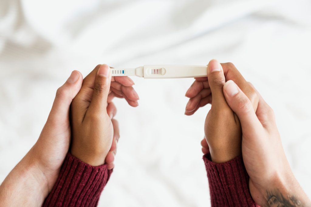
Pregnancy tests function by identifying the presence of a hormone known as human chorionic gonadotropin (hCG) in your urine. This hormone is produced by the cells that form the placenta, which nourishes the fertilised egg after it implants in the uterus. The level of hCG in your urine increases rapidly after conception, which is what pregnancy tests detect.

The Reliability of Home Pregnancy Tests

Home pregnancy tests are very reliable when used correctly. Most tests claim to be over 99% accurate. However, the accuracy of the test can vary depending on several factors, including the test’s sensitivity, how it is used, and when it is taken.

When to Take a Pregnancy Test

A few brands recommend taking a pregnancy test a week after you have missed your period. However, some of the latest test kits available in the market detect pregnancy as early as five days before your next period date. In addition, these early detection tests are reliable and are readily available over the counter.

Suppose you have irregular periods or do not keep track of your menstrual cycle. In that case, it may be more challenging to determine when to take a pregnancy test. In this case, it is best to wait at least three weeks after unprotected sex before taking a test.

Accuracy of Pregnancy Test Results

As mentioned earlier, home pregnancy tests are reliable when used correctly. However, it is essential to note that no test is 100% accurate, and there is always a chance of getting a false positive or negative result.

A false positive result is when a pregnancy test indicates that you are pregnant when you are not. On the other hand, a false negative result occurs when a pregnancy test suggests that you are not expecting a baby, even though you are.

Factors Affecting the Accuracy of Pregnancy Test Results

Several factors can affect the accuracy of pregnancy test results. These include:

  • The sensitivity of the test: Some tests are more sensitive than others and can detect lower levels of hCG in your urine. Tests that claim to detect pregnancy early may not be as reliable as tests taken after your missed period.
  • How the test is used: It is crucial to read and follow the instructions carefully when taking a pregnancy test. You may get an inaccurate result if you do not use the test correctly.
  • Medications: Certain medications, such as fertility drugs, can affect the levels of hCG in your urine and lead to a false positive result.
  • Medical conditions: Some medical conditions, such as ovarian cysts and ectopic pregnancy, can also affect the levels of hCG in your urine and lead to a false positive or false negative result.

Conclusion

In conclusion, home pregnancy tests are a convenient and reliable way to determine if one is pregnant. However, it is essential to take the test at the right time, follow the instructions carefully, and be aware of the factors that can affect the accuracy of the results. If you get a negative result but still suspect you may be pregnant, it is best to wait a few days and then take another test. Consulting with a healthcare professional is always a good idea if you continue to have questions or concerns.

FAQs

In conclusion, home pregnancy tests are a convenient and reliable way to determine if one is pregnant. However, it is essential to take the test at the right time, follow the instructions carefully, and be aware of the factors that can affect the accuracy of the results. If you get a negative result but still suspect you may be pregnant, it is best to wait a few days and then take another test. Consulting with a healthcare professional is always a good idea if you continue to have questions or concerns.

Q: How do pregnancy tests work?
A: Pregnancy tests work by detecting a hormone called human chorionic gonadotropin (hCG) in your urine. This hormone is produced by the cells that form the placenta, which nourishes the fertilised egg after it implants in the uterus. The level of hCG in your urine increases rapidly after conception, which is what pregnancy tests detect.

Q: When is the best time to take a pregnancy test?
A: A few brands recommend taking a pregnancy test a week after you have missed your period. However, some of the latest test kits available in the market detect pregnancy as early as five days before your next period date. These early detection tests are reliable and are easily available over the counter.

Q: How accurate are home pregnancy tests?
A: Home pregnancy tests are very reliable when used correctly. Most tests claim to be over 99% accurate. However, the accuracy of the test can vary depending on several factors, including the sensitivity of the test, how it is used, and when it is taken.

Introduction
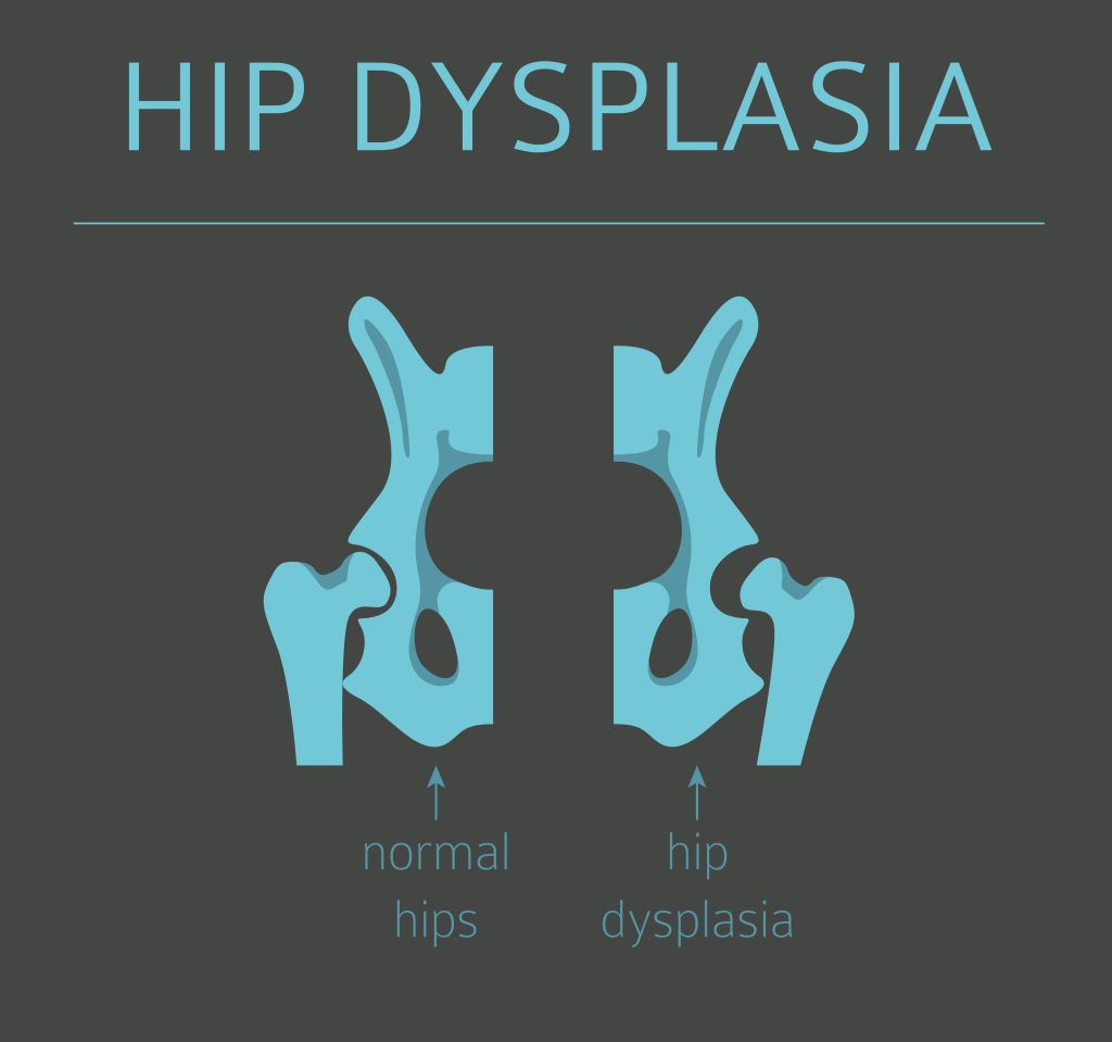
Hip dysplasia is a medical term for a common congenital condition that affects the hip joint, causing it to develop abnormally. The hip joint is critical for walking, running, and jumping. It is one of the largest weight-bearing joints. In hip dysplasia, the hip joint socket does not fully cover the ball of the upper thighbone, leading to instability and joint damage over time. This condition can cause significant discomfort and impact quality of life, especially if left untreated. With improper development, the hip joint becomes partially or completely dislocated, increasing the risk of arthritis and joint deterioration later in life. It’s a common condition and affects individuals of all ages, from infants to elderly adults. Those with hip dysplasia are typically born with this condition

Symptoms of Hip Dysplasia
The symptoms of hip dysplasia can vary based on the severity of the condition and by age group ranging from mild to severe. Some common symptoms include:

  • Pain or discomfort in the hip joint
  • Limping or favouring one leg over the other
  • Decreased range of motion in the hip joint
  • Popping or clicking sounds in the hip joint
  • A waddling gait or sway in the hip region

It’s important to note that some individuals experience no symptoms at all, thus, making it essential to receive regular check-ups to monitor the health of the hip joint. 
 In the case of infants, doctors check for signs of hip dysplasia shortly after birth and during follow-up visits. Usually, you will notice one leg is shorter than the other and find one hip less flexible during a diaper change. Or when the child starts walking, they may develop a limp. A soft brace usually corrects the problem of hip dysplasia if it is diagnosed in early infancy.
 In mild cases of hip dysplasia, symptoms may not start until a person is a teenager or young adult, damaging the cartilage lining the joint, and hurting the soft cartilage (labrum) lining the socket, causing a hip labral tear. It can result in activity-related groin pain and a sensation of hip instability. Surgery is usually recommended to move the bones into the proper positions for smooth joint movement.

Causes of Hip Dysplasia
The hip joint is formed from soft cartilage at birth, gradually becoming bone. The ball and socket must fit correctly as they shape each other. If the ball isn’t properly seated in the socket, the socket will be shallow and not fully formed around the ball. Crowding in the womb during the final month before birth can cause the hip joint ball to move out of place, leading to a shallower socket. The following factors may cause hip dysplasia- 

  • First pregnancy, 
  • Large baby, or 
  • Breech presentation.

Risks and Complications of Hip Dysplasia
Hip dysplasia has a genetic tendency and is more prevalent in girls. The risk of developing hip dysplasia increases in babies born in the breech position and those tightly swaddled with straight hips and knees. Hip dysplasia can increase the risk of developing other hip problems later in life, including arthritis and joint deterioration. These conditions can lead to significant pain and disability and may require surgery to correct. Hip dysplasia can also cause a person to develop a limp or walk with an awkward gait, leading to further musculoskeletal problems like arthritis and other long-term joint problems if left untreated.

Treatment Options for Hip Dysplasia
The treatment options for hip dysplasia may vary depending on the severity of the condition and the patient’s age. For children, treatment may include physical therapy and bracing to help improve hip alignment and strengthen the muscles surrounding the joint.
 For adults, treatment options may include non-surgical options such as physical therapy, weight management, and pain management. Surgery may be recommended in more severe cases to correct the hip joint and prevent future damage.

  • Prevention of Hip Dysplasia
  • There is no surefire way to prevent hip dysplasia. Still, some steps can reduce the risk of developing this condition. These include:
  • Maintaining a healthy weight
  • Engaging in regular physical activity to strengthen the muscles surrounding the hip joint
  • Avoiding high-impact activities that put excessive stress on the hip joint
  • Wearing proper footwear to help maintain proper hip alignment

Conclusion
Hip dysplasia is a common congenital condition that affects the hip joint and can lead to significant discomfort and disability if left untreated. Treatment options for hip dysplasia vary based on the severity of the condition and the patient’s age. They may include physical therapy, bracing, weight management, and surgery. By taking steps to maintain a healthy weight, engage in regular physical activity, avoid high-impact activities, and wear proper footwear, individuals can reduce their risk of developing hip dysplasia and maintain a high quality of life. You must have regular check-ups with a healthcare provider to monitor the health of the hip joint and address any issues that may arise.

FAQs 
 Q: What is hip dysplasia?
Hip dysplasia is a congenital condition in which the hip socket is shallow that does not fully cover the ball, causing instability in the joint and increasing the risk of arthritis and joint deterioration.
 
Q: Who is at risk for hip dysplasia?
Anyone can develop hip dysplasia, but it is common in individuals with a family history of the condition, women, and people with certain genetic conditions.
 
Q: What are the symptoms of hip dysplasia?
The symptoms of hip dysplasia can include pain or discomfort in the hip joint, limping or favouring one leg over the other, decreased range of motion in the hip joint, popping or clicking sounds in the hip joint, and a waddling gait or sway in the hip region.
 
Q: How is hip dysplasia diagnosed?
Hip dysplasia is typically diagnosed through a physical examination and imaging tests, such as X-rays or MRI scans.
 
Q: How is hip dysplasia treated?
Treatment options for hip dysplasia, depending on the severity of the condition and the patient’s age may include physical therapy, bracing, weight management, and surgery.
 
Q: Can hip dysplasia be prevented?
While hip dysplasia cannot be prevented, individuals can reduce their risk of developing the condition through weight management and regular physical activity, avoiding high-impact activities and wearing proper footwear.
 
Q: Can hip dysplasia cause long-term complications?
If left untreated, hip dysplasia can increase the risk of developing other hip problems later in life, including arthritis and joint deterioration, which can cause significant pain and disability.
 
Q: When should I see a doctor for hip dysplasia?
If you are experiencing symptoms of hip dysplasia, such as pain or discomfort in the hip joint or a limp or waddling gait, it is important to see a doctor. Regular check-ups with a healthcare provider can also help monitor the health of the hip joint and address any issues that may arise.

References :

What are Absorbable Sutures?

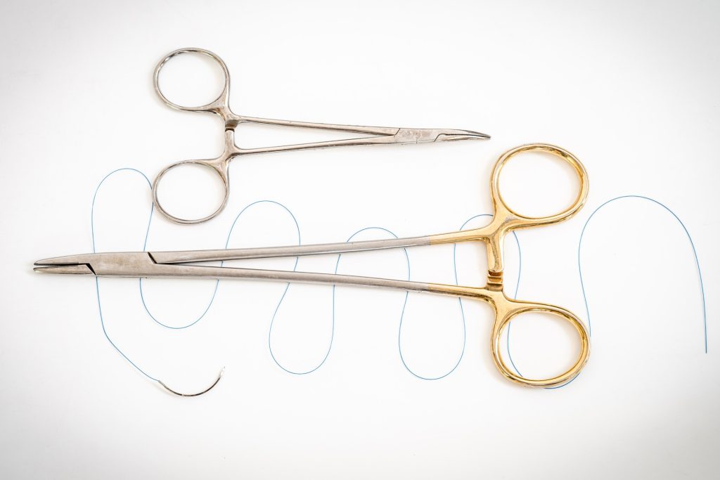
When undergoing surgery, sutures are necessary to close wounds and promote healing. Traditionally, non-absorbable sutures were used, requiring a follow-up appointment to remove them. However, with advancements in medical technology, absorbable sutures have become increasingly popular. 

Absorbable sutures are typically made from materials the body can naturally break down. As the body breaks down these materials, they are gradually absorbed and eliminated, leaving behind a healed wound and eliminating the need for removal. 

During surgery, the use of absorbable sutures is generally similar to that of non-absorbable sutures. The surgeon will close the wound using the suture, tying it off to ensure proper wound closure.

However, not all absorbable sutures are created equal. Different types of absorbable sutures have varying strengths and breakdown rates, which can impact their effectiveness in wound closure.

Types of Absorbable Sutures

Let’s discuss the different types of absorbable sutures and how they may vary in terms of strength and breakdown rate: 

  • Polyglactin (Mitsu) Sutures: Polyglactin sutures are popular for wound closure. They have excellent tensile strength and can maintain wound support for up to 42 days. They are made of a synthetic polymer designed to dissolve slowly and get absorbed by the body within 56 to 70 days.
  • Catgut Sutures: Catgut suture is a type of absorbable suture made from the sub mucosa of sheep or goat intestines. Traditionally, it has been used for centuries in various surgical procedures, but it is less common today due to the availability of synthetic absorbable sutures. Catgut suture is known for its rapid breakdown rate.
  • Poliglecaprone 25 (Filapron) Sutures: Poliglecaprone sutures are a type of absorbable sutures made of a synthetic polymer designed to break down quickly and be absorbed by the body within 90 to 120 days. They have excellent tensile strength and are perfect for procedures requiring Subcutaneous Layer Closure.
  • Polydioxanone (Filaxyn) Sutures: Polydioxanone sutures are another synthetic option designed to dissolve slowly over time. They are particularly useful for procedures that require extended wound support, as they can maintain tensile strength for up to 180 days. They are often used in surgeries that involve internal organs or tissues. Filaxyn suture is often used in surgeries that require extended wound support, such as internal organ or Fascia tissue repair.

Benefits of Absorbable Sutures

There are several benefits to using absorbable sutures. Some of them are as follows:

  • They are cost-effective and eliminate the need for a second surgery to remove the sutures, thus saving time and reducing the risk of complications.
  • Absorbable sutures are also less likely to cause an inflammatory reaction, which can help reduce the risk of infection and promote faster healing.
  • They offer excellent tensile strength and can maintain their integrity for an extended period, giving surgeons more time to complete the procedure without the sutures breaking or degrading.
  • Absorbable sutures are available in different materials, allowing surgeons to choose the most appropriate type for the specific procedure.
  • They are suitable for various surgical procedures, including orthopedic surgery, plastic surgery, and gynecological procedures.
  • Lastly, absorbable sutures may reduce scarring as they do not leave behind permanent suture marks.

Recovery and Healing with Absorbable Sutures

After surgery, the healing process with absorbable sutures is generally similar to that with non-absorbable sutures. Patients may experience swelling and pain, which can be managed with medication and other therapies. Additionally, patients may need to follow specific care instructions, such as changing wound dressings or avoiding certain activities that may strain the wound.

Following your surgeon’s post-operative care instructions is essential to ensure proper wound healing. While absorbable sutures eliminate the need for suture removal, they still require proper care to prevent infection and ensure proper healing.

Conclusion

In conclusion, absorbable sutures offer a range of benefits for both surgeons and patients. They eliminate the need for suture removal, reduce infection risk, and improve wound healing outcomes. However, as a patient, it is important to know the type of sutures your surgeon plans to use and what to expect during and after your procedure. By understanding the benefits and limitations of absorbable sutures, you can play an active role in your healthcare and achieve the best possible outcomes. 

FAQs

Q: How do absorbable sutures differ from non-absorbable sutures?
A: 
Absorbable sutures are designed to break down over time and are made from materials that can be naturally absorbed and eliminated by the body. On the other hand, non-absorbable sutures are not intended to be broken down by the body and require removal by a healthcare professional.

Q: How long does it take for absorbable sutures to break down?
A:
 The breakdown time for absorbable sutures will vary depending on the material type used. Some absorbable sutures may break down within a few weeks, whereas others may take several months.

Q: Do patients need to follow special care instructions after surgery with absorbable sutures?
A:
 Yes, patients will still need to follow specific care instructions after surgery with absorbable sutures to ensure proper wound healing. This may include changing wound dressings, avoiding certain activities, and taking prescribed medications as directed by their healthcare provider.

Reference links:

What Is Rectal Cancer?
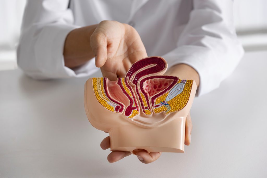
Rectal cancer develops in the rectum, a chamber between the end of the large intestine and the anus. It usually starts as a growth on the inner lining of the rectum and can spread to other body parts if left untreated. Symptoms of rectal cancer may include changes in bowel habits, rectal bleeding or blood in the stool, abdominal discomfort, and unexplained weight loss.

Diagnosis and Staging of Rectal Cancer
Diagnosing rectal cancer involves a combination of tests and procedures that help identify cancer cells in the rectum. Standard diagnostic tests include a digital rectal exam, colonoscopy, CT scan, MRI, and biopsy. These tests help determine the location and size of cancer and whether it has spread to nearby tissues or organs. In addition, the staging of rectal cancer is crucial in determining the most appropriate treatment options. The TNM staging system is used to stage rectal cancer based on tumour size, lymph node involvement, and distant metastases.

Treatment Modalities for Rectal Cancer in India
The treatment of rectal cancer typically involves a combination of surgery, radiation therapy, and chemotherapy. The choice of treatment modality depends on the cancer stage, the tumour’s location, and the patient’s overall health. The treatment modality includes- 

  • Surgery: Surgery is the most common treatment modality for rectal cancer. It is a highly effective treatment, mainly when the cancer is detected early. The benefits of surgery include the removal of cancerous tissue and the prevention of cancer recurrence. It involves the removal of the cancerous tumour along with surrounding tissues and lymph nodes. The type of surgical procedure used depends on the stage and location of cancer.
  • Radiation Therapy: Radiation therapy is a treatment modality that uses high-energy radiation to kill cancer cells. It is usually applied with surgery to kill any remaining cancer cells that may not have been removed during surgery. The benefits of radiation therapy include the destruction of cancer cells and the prevention of cancer recurrence.
  • Chemotherapy: As a treatment modality, chemotherapy uses drugs to kill cancer cells. It is typically used with surgery and radiation therapy for advanced-stage rectal cancer or when cancer has spread to other body parts. Chemotherapy drugs can be administered orally or intravenously. It can help shrink tumours, prevent cancer recurrence, and slow the progression of advanced-stage cancer. Chemotherapy can also improve the patient’s quality of life by relieving symptoms and reducing pain.

Choosing the Right Treatment Modality for Rectal Cancer
Choosing the suitable treatment modality for rectal cancer depends on several factors, including the cancer stage, the tumour’s location, the patient’s overall health, and preferences.

Conclusion
Rectal cancer is a significant health concern in India, and early detection and treatment are crucial for successful outcomes. With early detection and proper treatment, rectal cancer can be successfully treated, and patients can go on to live healthy and fulfilling lives.

FAQs
Q: What is the most common treatment modality for rectal cancer?
A:
 Surgery is the most common treatment modality for rectal cancer. Different surgical procedures may be used depending on the stage and location of the tumour.

Q: Can chemotherapy be a standalone treatment modality for rectal cancer?
A:
 Chemotherapy is rarely used as a standalone treatment modality for rectal cancer. It is usually used with other treatments, such as surgery and radiation therapy, to improve outcomes.

Q: Which factors should be considered when selecting a treatment modality for rectal cancer?
A:
 Several factors should be considered when selecting a treatment modality for rectal cancer, including the stage of cancer, the location of the tumour, the patient’s overall health, and the patient’s preferences and values.

Reference links:
https://www.cancercenter.com/cancer-types/colorectal-cancer/types/care-at-city-of-hope#:~:text=Rectal%20cancer%20occurs%20when%20cells,with%20colorectal%20cancer%20is%2068.
https://www.cancer.gov/types/colorectal/screening-fact-sheet
https://www.cancer.org/cancer/colon-rectal-cancer/treating/rectal-surgery.html

What is a cardiac valve?
The cardiac valves are structures in the heart that regulate blood flow through the heart’s chambers and into the major blood vessels. The cardiac valves are crucial for overall cardiovascular health. Problems with the valves can cause various health issues. Treatment options may include medication or surgery.

What is valve replacement surgery?
Valve replacement surgery is when a diseased or damaged heart valve is replaced with a new valve. Several types of valves can be replaced, like the aortic valve, mitral valve, tricuspid valve, and pulmonary valve. The specific procedure used to replace a valve will depend on the location, the type of valve being replaced, and the patient’s overall health.

Importance of Sutures in Valve Replacement Procedure
The significance of sutures in valve replacement procedures cannot be overstated. Their importance is stated as follows:

  • Secure Replacement Valve in Place: Sutures are used to secure the replacement valve in place once it has been implanted. The suture material attaches the replacement valve to the surrounding tissue. It must be strong and secure, so the replacement valve stays in place and functions properly.  The suture material is usually passed through the surrounding tissue and then tied off, creating a secure connection between the replacement valve and the surrounding tissue. Hence, the suture material must be strong enough to withstand the forces exerted on the valve during normal cardiac function and be flexible enough to allow the valve to close and open properly. 
  • Promotes Healing: Surgical sutures also play an important role in promoting healing after a valve replacement procedure. They help close the incision made during the surgery. Their placement and technique are essential in ensuring that the scar is minimal and as inconspicuous as possible. However, the type of suture used can also impact healing time and the risk of complications.
  • Prevention of Complications: Each valve replacement requires a specific suture and suture technique to avoid risks and complications. Properly placed and tightened sutures can help minimize complications such as blood clots, heart failure, a periprosthetic leak, prosthesis-patient mismatch, and surgical site infections.

Importance of Suture Techniques and Materials in Valve Replacement Procedure

Suture Techniques
Multiple suture techniques may be used in valve replacement, including:

  1. Interrupted Suturing: In this technique, individual sutures are used to secure the prosthetic valve to the surrounding tissue. The sutures are placed at regular intervals along the circumference of the valve annulus, ensuring a secure attachment.
  2. Continuous Suturing: Continuous suturing involves the use of a single suture thread that is passed through the valve annulus in a continuous manner. This technique provides a more secure and watertight closure compared to interrupted suturing.
  3. Modified Continuous Suturing: This technique is a variation of continuous suturing. Instead of using a single continuous suture, multiple short segments of suture thread are used. This allows for better control and adjustment of tension at various points around the valve annulus.
  4. U-Shape Suturing: In this technique, U-shaped sutures are used to secure the prosthetic valve. The sutures are passed through the valve annulus and tied on either side, creating a U-shape that holds the valve in place.
  5. Horizontal Mattress Suturing: This technique involves the use of sutures that pass through the valve annulus in a horizontal direction, creating a “mattress” effect. This technique provides good tissue approximation and helps distribute tension evenly.

Apart from these the major suturing technique that is used most often for suturing is the Pledgeted Suturing Technique

The pledgeted suturing technique is commonly used in valve replacement surgery to reinforce the suture line and provide added security. Pledgets are small pieces of felt or Teflon-like material that are placed on the sewing ring of the prosthetic valve. These pledgets act as a cushion between the suture and the delicate tissue of the heart, helping to distribute the suture tension more evenly and reduce the risk of tearing or tissue damage.
Here’s how the pledgeted suturing technique is typically performed in valve replacement surgery:

  1. After the diseased valve is removed and the annulus (the ring of tissue where the valve sits) is prepared, the prosthetic valve is positioned in place.
  2. The surgeon uses sutures (commonly non-absorbable sutures like braided polyester) to secure the valve to the annulus. These sutures are typically passed through the sewing ring of the prosthetic valve and then through the tissue of the annulus.
  3. Prior to tying the suture, a pledget is placed on the ventricular (lower) side of the annulus. The pledget is positioned between the annulus tissue and the suture.
  4. The suture is then tied, securing the prosthetic valve in place. The pledget acts as a protective layer between the suture and the heart tissue, minimizing the risk of tearing or cutting into the tissue.

The pledgeted suturing technique helps to provide a more secure attachment of the prosthetic valve while reducing the potential for damage to the delicate heart tissue. By distributing the tension more evenly, it can help improve the longevity and durability of the valve replacement.

Suture Materials

The choice of suture material depends on several factors, including the type of tissue being sutured, the surgeon’s preference, and the specific requirements of the surgery. A cardiac surgeon will determine the most appropriate suture material based on these considerations.

Currently the industry standard for the suture material being used for valve replacement is Polyester. Usually a braided polyester suture with a ½ circle Taper cut Needle in a combination of green and white colors and PTFE pledgets is used for securing the prosthetic valve in place. It has excellent handling characteristics and also provides good knot security.

Dangers of Choosing the Wrong Kind of Suture Material and Technique

Choosing the wrong suture material for valve replacement surgery can potentially lead to various complications and adverse outcomes. Here are some dangers associated with using an inappropriate suture material:

  1. Suture Breakage: If an insufficiently strong suture material is used, there is a risk of suture breakage, particularly in high-stress areas. This can result in the prosthetic valve becoming loose or dislodged, compromising its function and potentially leading to valve failure.
  2. Suture Degradation or Absorption: Using an absorbable suture material in valve replacement surgery can be problematic. Absorbable sutures are designed to break down and be absorbed by the body over time. However, in valve replacement surgery where long-term durability is crucial, the suture material needs to remain intact and provide long-lasting support. Absorbable sutures may degrade prematurely, leading to valve instability or failure.
  3. Tissue Damage and Suture Cutting: Inadequate suture material can cause tissue damage, especially if it is too sharp or lacks proper cushioning. This can result in tissue tearing or cutting, leading to bleeding, leakage around the suture line, or compromised tissue integrity.
  4. Infection Risk: The choice of suture material can influence the risk of post-operative infections. Certain suture materials may be more prone to harboring bacteria or promote bacterial adherence, increasing the likelihood of surgical site infections. This can have detrimental effects on the healing process and overall patient outcomes.
  5. Tissue Irritation or Reaction: Inappropriate suture materials may cause tissue irritation, inflammation, or allergic reactions in some individuals. This can impede proper healing, lead to discomfort or pain, and potentially require additional interventions for resolution.

To minimize these risks, it is essential for the surgeon to carefully select the appropriate suture material based on factors such as tissue type, surgical technique, patient characteristics, and the specific requirements of the valve replacement procedure. Close adherence to established surgical guidelines and the expertise of the surgical team are vital in ensuring the use of the most suitable suture material to achieve optimal outcomes in valve replacement surgery.

Conclusion

With all the benefits of a new valve and increased longevity, it is no wonder that people are eager to undergo a cardiac valve replacement procedure. However, it is important to choose the correct procedure for you. Ensure you get proper preoperative and postoperative care for the best outcome from your treatment. Early consultation with a cardiac surgeon is the best way to determine which procedure is best for you. Suppose you are considering a valve replacement procedure. In that case, you should be aware of the steps involved in the procedure and the types of valves available.

FAQs
 
Q. What is the importance of surgical sutures in valve replacement procedures?
A.
 Surgical sutures are used to secure the replacement valve and ensure it functions properly. The suture material attaches the new valve to the surrounding tissue. Therefore, it must be strong and secure to withstand the forces exerted on the valve during normal cardiac function. Additionally, sutures play a crucial role in promoting healing after the surgery, helping to close the incision made during the procedure and minimize scarring.
 
Q. What are cardiac valves, and why are they essential for overall cardiovascular health?
A.
 Cardiac valves are structures in the heart that regulate blood flow through the heart’s chambers and into the major blood vessels. There are four main types of cardiac valves: the aortic valve, mitral valve, tricuspid valve, and pulmonary valve. These valves are essential for overall cardiovascular health because they ensure that blood flows through the heart in the correct direction, preventing backflow and ensuring all body parts receive the oxygen and nutrients they need.

Q. What are the different types of suture material that can be used in valve replacement procedures, and how are they chosen?
A.
 Currently the industry standard for the suture material being used for valve replacement is Polyester. Usually a braided polyester suture with a ½ circle Taper cut Needle in a combination of green and white colors and PTFE pledgets is used for securing the prosthetic valve in place. It has excellent handling characteristics and also provides good knot security.

Q. Which suture techniques may be used in valve replacement procedures, and how do they differ?
A.
 The pledgeted suturing technique is commonly used in valve replacement surgery to reinforce the suture line and provide added security. Pledgets are small pieces of felt or Teflon-like material that are placed on the sewing ring of the prosthetic valve. These pledgets act as a cushion between the suture and the delicate tissue of the heart, helping to distribute the suture tension more evenly and reduce the risk of tearing or tissue damage.
Here’s how the pledgeted suturing technique is typically performed in valve replacement surgery:

  1. After the diseased valve is removed and the annulus (the ring of tissue where the valve sits) is prepared, the prosthetic valve is positioned in place.
  2. The surgeon uses sutures (commonly non-absorbable sutures like braided polyester) to secure the valve to the annulus. These sutures are typically passed through the sewing ring of the prosthetic valve and then through the tissue of the annulus.
  3. Prior to tying the suture, a pledget is placed on the ventricular (lower) side of the annulus. The pledget is positioned between the annulus tissue and the suture.
  4. The suture is then tied, securing the prosthetic valve in place. The pledget acts as a protective layer between the suture and the heart tissue, minimizing the risk of tearing or cutting into the tissue.

The pledgeted suturing technique helps to provide a more secure attachment of the prosthetic valve while reducing the potential for damage to the delicate heart tissue. By distributing the tension more evenly, it can help improve the longevity and durability of the valve replacement.

Q. Which type of suture is the best for cardiac valve replacement surgery?
A.
 Polyester Sutures are considered the best for Valve replacement surgery as they provide excellent handling characteristics and good knot security.