Category

Peripheral Vascular

Category

Have you ever felt like stress is taking a toll on your health? You’re not wrong. Stress affects more than just your mind; it can have severe consequences on your body, too, especially when it comes to peripheral vascular disease (PVD). Peripheral vascular disease, a condition where narrowed arteries reduce blood flow to the limbs, can be worsened by chronic stress. But how exactly does stress impact this condition, and what can you do to manage it? Let’s explore the connection between stress and peripheral vascular disease and discover practical ways to improve your vascular health through effective stress management.

How Stress Affects Vascular Health

Stress responses in the body are natural reactions designed to protect us in dangerous situations. However, chronic stress might lead to various health problems, including peripheral vascular disease. When we experience stress, our bodies tend to release hormones such as adrenaline and cortisol. These hormones prepare the body for a “fight or flight” response, leading to an increase in heart rate and blood pressure[1]. While this response is beneficial in short bursts, prolonged stress can cause significant harm to vascular health.

Stress Responses

When stress triggers the release of adrenaline and cortisol, it causes the blood vessels to constrict, leading to higher blood pressure. This increased pressure on the vascular system can damage the inner lining of the arteries. This makes them more susceptible to plaque buildup[2]. Over time, this can contribute to the development and worsening of peripheral vascular disease.

Long-term Effects

Chronic stress can lead to persistent inflammation in the body[3], which is a risk factor for peripheral vascular disease. Inflammation can cause damage to the vascular walls. This in turn promotes plaque formation and increases the risk of thrombosis (blood clots). These clots might obstruct blood flow and lead to complications such as heart attacks or strokes.

Stress and PVD Risk

Several studies have highlighted the link between stress and an increased risk of developing peripheral vascular disease. Individuals experiencing high levels of stress are more likely to develop PVD compared to those with lower stress levels[4]. This underscores the importance of effective stress management strategies to prevent and manage peripheral vascular disease.

Stress Triggers and Symptoms in PVD

Understanding the triggers and symptoms of stress in individuals with peripheral vascular disease is crucial for effective management. Stressors can be both emotional and physical, each contributing to the worsening of PVD.

  • Emotional stressors such as job pressure, family issues, and financial worries can significantly impact vascular health[5]. These stressors lead to increased cortisol levels, promoting inflammation and vascular damage.
  • Physical stressors, including sleep deprivation and poor diet, can also worsen the symptoms of peripheral vascular disease[6][7]. Lack of sleep can increase blood pressure, while a poor diet can lead to the accumulation of unhealthy fats in the blood vessels.

Symptoms of Peripheral Vascular Disease to Watch For[8]

Individuals with peripheral vascular disease should be aware of the symptoms that might indicate increased stress levels affecting their condition.

  • Intermittent claudication, characterized by pain and cramping in the legs during physical activity, is a common symptom.
  • Changes in skin colour, such as paleness or a bluish tint, can also indicate poor blood flow.
  • Additionally, non-healing ulcers or sores on the legs and feet are critical signs that stress may be worsening the condition.

Managing Stress to Improve Vascular Health[9]

Effectively managing stress is important for improving vascular health and preventing the progression of peripheral vascular disease. Several strategies can help reduce stress and its impact on vascular health.

  • Exercise: Regular low-impact exercises like walking, swimming, and cycling reduce stress and improve cardiovascular health.
  • Diet: A diet rich in antioxidants, whole grains, fruits, and vegetables reduces inflammation. Avoid excessive salt, oily & processed foods.
  • Mindfulness and Relaxation: Incorporate meditation, yoga, and deep breathing to reduce stress and promote relaxation.
  • Professional Help: Seek therapy for chronic stress. Cognitive-behavioural therapy offers effective coping strategies.
  • Healthcare Providers: Consult healthcare providers for personalized stress reduction and treatment adjustments for optimal vascular health.

Wrapping It Up

Stress plays a significant role in the progression of peripheral vascular disease. Understanding the impact of stress and implementing effective management strategies can significantly improve outcomes for individuals with PVD.

[1] https://www.health.harvard.edu/staying-healthy/understanding-the-stress-response
[2] https://www.urmc.rochester.edu/encyclopedia/content.aspx?ContentTypeID=1&ContentID=2171#:~:text=Studies%20suggest%20that%20the%20high,plaque%20deposits%20in%20the%20arteries.
[3]https://www.yalemedicine.org/news/stress-affects-your-heart#:~:text=%E2%80%9CLong%2Dterm%E2%80%94or%20chronic,heart%20rhythms%2C%20and%20heart%20failure.
[4]https://newsroom.heart.org/news/work-related-stress-linked-to-increased-risk-for-peripheral-artery-disease#:~:text=Researchers%20found%20that%20people%20with,age%2C%20sex%20and%20lifestyle%20variables.
[6] https://www.medicalnewstoday.com/articles/insufficient-sleep-tied-to-increased-risk-of-a-condition-that-may-lead-to-stroke#Using-genetic-data-to-study-PAD-risk
[8]https://www.betterhealth.vic.gov.au/health/conditionsandtreatments/peripheral-vascular-disease
[9] https://www.urmc.rochester.edu/encyclopedia/content.aspx?ContentTypeID=1&ContentID=2171#:~:text=Get%20plenty%20of%20exercise&text=Exercise%20can%20help%20to%20improve,reduced%20physical%20response%20to%20stress.
https://www.mayoclinic.org/diseases-conditions/high-blood-pressure/in-depth/stress-and-high-blood-pressure/art-20044190

https://www.verywellmind.com/what-is-the-fight-or-flight-response-2795194#:~:text=These%20hormones%20cause%20the%20sympathetic,blood%20pressure%2C%20and%20breathing%20rate
.
https://newsroom.heart.org/news/work-related-stress-linked-to-increased-risk-for-peripheral-artery-disease
https://www.betterhealth.vic.gov.au/health/conditionsandtreatments/peripheral-vascular-disease

Introduction

Experience of any sharp pain or numbness in your legs after an extended period of sitting, painful cramps or aching in your hips, thighs, or calf muscles common and frequent after any physical activity, or coldness in lower feet, are signs of blockages in the arteries of the lower extremities. The condition is known as peripheral artery disease. As per reports, it is common, with nearly 41 to 54 million[1] estimated cases in India and nearly 4.2.to 6.2 million[1] people are afflicted by critical limb ischemia. According to the American Heart Association statement, approx. two hundred million people suffer from arterial blockages of the extremities worldwide.

The alarming statistics and the rising cases of other complications associated with arterial blockages in diabetic patients and people with smoking and alcohol consumption and/or obesity raise more significant concern for its stringent preventive measures and prompt and effective address.

The blog discusses the condition of arterial blockages, its causes, symptoms, and the treatment options for its effective control and management.

Peripheral Artery Disease- Its Causes and Symptoms 

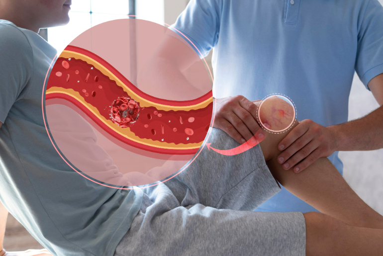
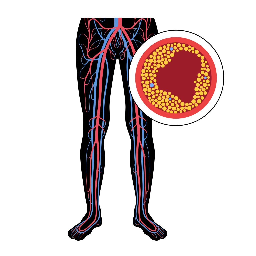
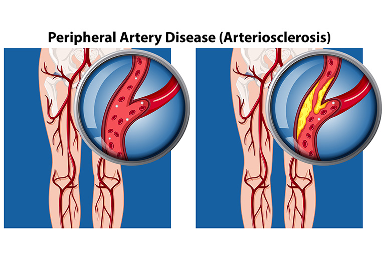
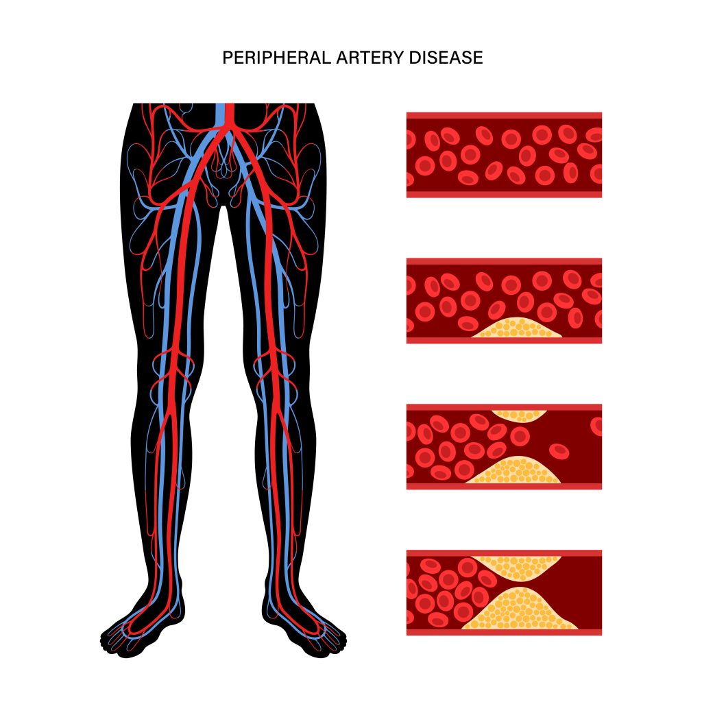
Peripheral Artery Disease (PAD) results from the narrowing of the arteries that carry blood from the heart to the peripheral arteries as well extremities, commonly the legs. The lack of oxygenated blood supply due to the narrowing of the arteries causes ischemic pain. The causes of arterial blockages are-

  • Atherosclerosis – Plaque (fatty deposits) buildup over time results in the narrowing of the arteries, interrupting the blood supply to the peripheral arteries.
  • Thrombosis– A clotting of the blood in the narrowed artery is one of the reasons for arterial blockages.
  • Embolism– A condition of blocked artery caused by a blood clot or an air bubble in which the smooth flow of blood to the artery is interrupted, leading to an insufficient supply of oxygen. This causes pain, cramps, and other related symptoms of PAD.

Symptoms of PAD

People with PAD may not experience any symptoms until they become severe, which could at times adversely affect the heart health of the patient, if left unaddressed. This is so because PAD shares many common risk factors like diabetes, high cholesterol, high blood pressure, obesity, and smoking with heart disease.

The main symptom of PAD is claudication, where the person experiences flared cramping and pain in the legs and thighs when walking and subsides when at rest. The other symptoms include coldness in feet, numbness, tingling, or slow healing or unhealing of wounds.

Treatment for PAD

Preventing diseases and related complications is the best way to live a healthy life and enrich your quality of life. People with PAD are at a high risk of cardiovascular diseases and chances of stroke, heart attack, and other heart-related issues. Treatment for PAD requires rigorous and stringent healthcare precautions and aggressive lifestyle modifications.

Self-care and medications

A healthy diet, regular exercise, quitting smoking, management and control of comorbidities through medications, effective stress management, regular checkups, and monitoring through routine tests go a long way in preventing PAD and other related complications.

Surgical and non-surgical approaches

After the color doppler test, ankle-brachial index and vascular ultrasound test, the healthcare provider decides whether the patient needs surgical intervention to treat PAD. The severity of the condition is the primary and deciding factor for surgical intervention with other anatomical and health conditions of the patient.

Vascular Surgery

Known as open surgery or bypass surgery, the surgeon creates a new path around the blocked artery, using the healthy blood vessels from the other part of the patient’s body. This is known as grafting. The blood flows through the new path, bypassing the blocked artery.

During this bypass surgery, the patient is put on general anesthesia to keep them comfortable. The surgeon makes incisions to access the arteries and veins. The surgeon may use healthy veins like the saphenous veins (superficial veins in the legs), cephalic veins (veins from the arm), femoral veins (from thighs in case of more extensive artery grafting) from the patient’s body or use a synthetic material for grafting when natural healthy veins cannot be used. The surgeon sews the bypass in place and closes the incisions.

Endovascular Interventions

Atherectomy- It is a minimally invasive procedure to remove plaque from blocked arteries. During the procedure, the healthcare provider uses a sharp blade or laser attached to the tip of the catheter to cut or scrape out the hard plaque. The atherectomy could be Excisional (the blade cuts the plaque in one direction), Orbital (the plaque gets removed with a spinning tool), Rotational (the blade cuts the plaque in a rotating movement), or Laser ablation (the plaque is removed using laser) atherectomy.

Angioplasty – To treat any blood vessel damage, the interventional healthcare professional accesses them through minimal invasiveness. In endovascular intervention, the healthcare professional usually makes a tiny incision in the groin to gain access to the damaged arteries. Using the X-ray real-time images, the catheter, with a balloon attached, is guided to the damaged artery. Once in place, the balloon is inflated, pushing the plaque to the sides of the arterial walls, and paving the way for the uninterrupted flow of the blood.

In the case of angioplasty, the healthcare professional implants a stent, clearing the passage for the blood to flow in the affected artery. A stent, a small mesh tube, keeps the artery open for blood flow. Once the stent is placed, the balloon is deflated, the catheter is removed, and the incision is closed.

Apart from treating peripheral arteries, these stents are widely used to treat various artery blockages, like the coronary, renal, and carotid arteries.

Angioplasty has been gaining acceptance as a standard of care for treating blocked arteries due to its minimal invasiveness, resulting in less pain and tissue scarring, faster healing and recovery, shorter hospital stays, and quicker return to normalcy.

Conclusion

There are many risk factors common between PAD and heart disease; hence, effective control and management of one help to prevent the incidence of the other. PAD with other conditions of diabetes, high blood pressure, high cholesterol, or obesity puts the patient at a higher risk of heart disease. Rigorous, consistent, and aggressive lifestyle changes are a must for its prevention. However, in severe cases, where self-care, medications, lifestyle changes, and other alternate treatment modalities do not offer the desired results, surgical and non-surgical treatment options should be resorted to for timely and effective treatment.

The choice of the medical treatment option depends on the healthcare professional’s decision, arrived at after considering the severity of the condition, the age, anatomy, and overall health of the patient. Surgical and non-surgical treatment help repair the damage to the artery by removing the blockages. However, one must own responsibility toward one’s health and adhere to the doctor-recommended diet plan, exercise regime, and recovery plans, and quit smoking and tobacco consumption post-treatment for healthy living.

References

https://journals.lww.com/cmii/fulltext/2020/18030/endovascular_therapy_in_vascular_surgery___how.2.aspx#:~:text=41%E2%80%9354%20million%2C%20with%20an%20estimated%204.2%E2%80%936.2%20million%20patients%20of%20critical%20limb%20ischemia
https://www.heart.org/en/news/2023/06/15/routine-tests-may-reduce-disparities-in-peripheral-artery-disease-and-amputation-risk
https://www.narayanahealth.org/blog/what-is-peripheral-arterial-disease-pad/#:~:text=According%20to%20an%20International%20organization,built%20up%20in%20the

Introduction

Arterial blockages are a slow and gradual narrowing of the blood vessels that carry oxygenated blood throughout the body. These blood vessels could be coronary (supplying blood to the heart), carotid (supplying blood to the brain and the head), peripheral (supplying blood to the body other than heart and brain), or renal (supplying blood to the kidneys) arteries that get blocked due to the accumulation of plaque (fibrous and fatty deposit) over time. This condition of plaque buildup, known as atherosclerosis, is the underlying condition in most arterial blockages. Also known as stenosis, blocked arteries hamper the regular smooth flow of oxygenated blood in the body, causing angina, claudication, kidney failure, high blood pressure, stroke, and other complications depending on the arteries affected by atherosclerosis.

Peripheral arterial disease (PAD) is one of the most common diseases with approx. two hundred million adults reportedly suffer from it, and the incidence increases to nearly 20% in older adults above 70 years. The disease affects the arteries of the extremities, often resulting in restricted mobility of the legs and the arms. The management and treatment for PAD starts with lifestyle changes, cardiovascular risk management, medications, alternate treatment modalities, or surgical or endoscopic treatment.

Continue reading this blog to know what to expect after peripheral artery stent replacement.

Peripheral Arterial Disease (PAD)

Atherosclerosis is the most common cause of PAD; the other causes may also include injury, inflammation, or exposure of the blood vessels to radiation therapy. The arteries of the legs and arms experience poor blood supply, causing symptoms of claudication (pain in the legs while walking and relief when at rest), and in extreme and severe cases, lead to gangrene.

People with diabetes, family history of PAD, smoking habits, obesity, high blood pressure, and high cholesterol are highly prone to developing the symptoms of PAD.

Lifestyle changes through a healthy diet, regular exercise, quitting smoking, stress management, and staying active helps control diabetes, blood pressure, cholesterol, and obesity, the contributing risk factors to PAD.

The treatment therapies aim to reduce the risk of PAD and improve mobility through exercises and walking. Patients less responsive to other therapies and lifestyle changes may need endovascular intervention

The procedure with a minimally invasive approach opens the blocked arteries by pushing the plaque to the arterial walls and placing the stent in the treated arteries. The procedure allows the smooth supply of blood to the extremities. Stents have been used for treating arterial blockages with high success and acceptance rates. Stents do not cure atherosclerosis. They treat the condition and relieve the patient from pain, discomfort, immobility, and dependence. Post-procedural care and management of the disease is a must for healthy living. Endovascular treatment with stent placement relieves the patient from the pain, inflammation, and discomfort associated with blocked peripheral arteries.

Alternatively, patients suffering from PAD can undergo a surgical treatment where a vascular surgeon cuts open the affected area and performs the procedure to cure the patient.

Life after Stent Placement 

Stents are a boon for people who are misfit for surgery and/or non-responsive to other treatment options. The stent placement as a percutaneous procedure offers lesser pain, faster healing, fewer tissue scars, faster recovery, shorter hospital stay, and sooner resumption to routine.

The life of the patients undergoing stent placement for peripheral arterial blockages changes post-treatment. They experience significant relief from the symptoms of PAD. They can live a life of their choice once they have completely recovered. Life becomes relatively active and enriching, with people able to manage their daily chores comfortably and without stress and pain. They regain a sense of worthiness and purpose as they are less dependent on others for their daily needs.

However, the comfort and ease of living a life require post-operative care and management. One must pay attention to the fact that prevention is the only cure. The life of an individual after stent placement may seem like below.

  • Immediately upon discharge from the hospital, the patient requires proper rest and downtime with physical activities for complete healing.
  • Wound care (Diabetic patients) becomes a priority in order to prevent infection and other complications.
  • Healing and recovery are faster with stent placement; compared to surgery, hence one is expected to resume routine in a week or two.
  • One must adhere to positive lifestyle changes to prevent its relapse.
  • With improved blood flow in the extremities, the patient feels less pain and more comfort in walking and staying active.
  • The need for regular follow-up with the doctor and routine medications may arise is mandatory to keep a check on the condition.

Conclusion

PAD is a progressive condition, often a sign of poor cardiovascular health. The symptoms may or may not be noticeable earlier. For the treatment for PAD to be successful and effective, one has to take ownership for one’s health by being consistent in living a healthy life through positive lifestyle choices.

Life resumes normalcy faster after stent placement than open surgery. The relief from pain and discomfort makes life enjoyable and fulfilling. The benefits of stent placement for PAD are multiple, provided one takes precautions with wound care (diabetic patients), regular follow-up visits with doctors, a healthy diet, regular exercises, excessive physical stress to the legs and the arms, controlling risk factors, and a positive mindset.

References

Peripheral Arterial Disease – StatPearls – NCBI Bookshelf (nih.gov)

Introduction

Thrombosis comes from the Ancient Greek – thrómbōsis, which means ‘clotting.’ It is the process of the formation of a blood clot inside a blood vessel and is also known as a thrombus. In the process, it causes blocking and obstructs blood flow in the affected area resulting in complications, especially if the clot moves to a critical part of the circulatory system, the brain, or the lungs. 

In the event of an injury of a blood vessel, either a vein or an artery, it is very typical for the human body to prevent further blood loss by using thrombocytes or platelets and fibrin to form a blood clot. When a piece of the blood clot or the clot breaks free, it can form an embolus that moves around the body in the bloodstream, obstructing blood flow to critical organs like the brain or the lungs, resulting in reduced oxygen supply and blood flow with severe complications.

Types of Thrombosis

Now that you know thrombosis occurs when blood clots block blood vessels, you will discover that there are two broad classifications of thrombosis depending on the type of blood vessel affected (arterial or venous) and the exact location of the blood vessel or the organ supplied by it. 

Venous Thrombosis – When thrombosis occurs in the veins or the blood clot blocks a vein, it is called venous thrombosis, also known as DVT, which stands for deep vein thrombosis. In this case, there is a blood clot in the affected part of the body, like the deep veins, axillary or subclavian veins (deep veins of the upper limb), hepatic veins (veins that drain venous blood from the liver), renal vein (the major blood vessel carrying blood from the kidney and the ureter), or jugular veins (veins located within the carotid sheath on either side of the neck). Veins typically from different parts of body to the heart.

Arterial thrombosis – When thrombosis occurs in the arteries, and the blood clot blocks an artery, it is known as arterial thrombosis. Arteries carry oxy-rich blood from the heart to the rest of the body. In the case of arterial thrombosis, the blood supply can be affected and restricted, which can further damage the tissue supplied by that particular artery. 

For example, if arterial thrombosis occurs in a blood vessel in the brain, or what we call arterial embolism, it can lead to a stroke. Similarly, the blood clots could migrate downstream and pose a risk affecting any organ. And if arterial thrombosis occurs in the arteries that supply blood to the heart muscle (coronary arteries), it can lead to a heart attack. The blood clots happen due to the hardening of the arteries or thickening of the artery walls, a condition called atherosclerosis in which there is a buildup of plaque or fatty deposits. This plaque buildup can lead to a rupture followed by a blood clot. Thrombus in the coronary artery can also cause myocardial infarction leading to ischemia. The reduced oxygen supply to the heart cells can often lead to cell death or necrosis. 

Causes of Thrombosis

  • Venous thrombosis can be caused by-

– Immobility

– Any injury or disease to the leg veins

– A fracture

– Being overweight or obese

– Certain medications that can increase the risk of clotting

– Genetic disorders

– Autoimmune disorders.

  • Arterial Thrombosis, on the other hand, is caused by the hardening of the arteries, called arteriosclerosis. This typically occurs when fatty or calcium deposits make the artery walls thick, leading to a fatty buildup of plaque which can burst to cause a rupture, followed by a blood clot.

Generally speaking, some people are predisposed to developing thrombosis and thromboembolism (like newborn babies in the neonatal phase). At the same time, some have a higher risk of developing either venous thrombosis or arterial thrombosis. Also, the chances of development of this condition of thrombosis increase with age and depend on several risk factors and lifestyle factors like the ones discussed above and the following, 

● Heart Condition

● Lung Condition

● Crohn’s Disease (an inflammatory bowel disease affecting the lining of the digestive tract)

● Major Fractures, burns, or injuries to the deep veins in the legs, arms, or the pelvis area

● Lack of movement and physical activity due to travel without moving, medical procedures like surgery or hospitalization, or leg paralysis

● Certain medications like birth control medicines and hormonal contraceptives which contain estrogen or chemotherapy drugs

● Any autoimmune or inflammatory disorder that promotes easy blood coagulation and blood thickening

● Any inherited gene conditions like a family history of blood clots, stroke, or heart attack

● Diet

FAQs:

What is a major cause of thrombosis?

The major cause of thrombosis could be any of the following,

– immobility

– any injury or disease to the leg veins

– a fracture

– being overweight or obese

– certain medications that can increase the risk of clotting

– Genetic disorders

– Autoimmune disorders.

What are the three factors that lead to thrombosis?

Any of the following can be risk factors for thrombosis,

– high blood pressure

– high cholesterol

– diabetes.

What is the most common thrombosis?

The most common thrombosis is arterial thrombosis, which happens in arteries that carry blood from the heart to the rest of the body, often considered to be one of the key reasons for heart attacks and strokes.

What are the signs of thrombosis?

The visible signs and symptoms of thrombosis are pain, swelling, and tenderness in one of the legs, which is usually the calf or thigh, a dull and heavy ache in the affected area, also flushing warm skin in the area of the clot, visibly red skin at the area below the knee and back of the leg.

Introduction

Blood vessels responsible for transporting blood throughout the body are the components of our circulatory system. Arteries, veins, and capillaries are the various blood vessels, each performing a specific function to regulate blood circulation, ensure the proper supply of oxygen and nutrients, and removal of waste from the body. Arteries supply oxygenated blood from the heart to different parts of the body, while veins return the deoxygenated blood from the rest of the body to the Heart. The capillaries act as a connection between the arteries and the veins.

Any blockage affecting the smooth blood flow in these blood vessels leads to various health problems relating to the heart, brain, and other body parts. Blood circulation disorder affecting the blood vessels and blood supply to –

  • The heart is called Coronary Artery Disease
  • The brain is called Cerebrovascular Disease
  • The body parts other than the heart and the brain is called Peripheral Vascular Disease.

In this article, we will discuss Peripheral Vascular Disease and know the causes, symptoms, types, and treatment of the diseases.

What is Peripheral Vascular Disease?

Peripheral Vascular Disease is the condition of blockage and narrowing of the arteries and veins in the legs, arms, and body parts other than the heart and the brain. Peripheral Artery Disease (PAD) and Peripheral Vascular Disease (PVD) are often used interchangeably. However, there is a difference between the two. PAD is the narrowing of the arteries due to the accumulation of plaque (i.e., fats and cholesterol) on the inner walls of the arteries that impair the blood flow from the heart to different parts of body, which remain deprived of oxygen and nutrients. While PVD refers to the blockage and narrowing of the arteries and veins in parts of the body other than the heart and the brain due to various reasons, including the accumulation of plaque. Thus, PVD is an umbrella term.

Causes of PVD

The primary cause of PVD is Atherosclerosis. The build-up of plaque, i.e., accumulation of fats and cholesterol, can be in any artery in the body. The fatty substances and cholesterol that block the arteries and thereby reduce the blood flow are known as Atheroma. The condition of this plaque formation in the arteries is called Atherosclerosis. Due to narrowing, when the arteries cannot supply enough blood to the legs, it causes pain, cramps, aches, and discomfort during physical activity, but the same subsides with rest. This condition of pain in the legs is called claudication. Atherosclerosis can happen in any body part. But it may be noted that Atherosclerosis is caused in the arteries and not in the veins.   

Besides, Atherosclerosis, the other responsible factors for PVD can be-

  • Coronary Artery Disease – people with blocked arteries of the heart are at risk of developing PVD.
  • Lifestyle – Lifestyle choices like smoking, sedentary living, poor eating habits, and drug use also play a significant role in developing PVD.
  • Medical conditions– Medical conditions include diabetes, high blood pressure, high cholesterol, obesity, and kidney disease, which may also be the reason for one developing PVD.
  • Injury – Injury to the legs and arms may cause the veins to narrow or swell, impairing blood flow from the legs or arms to the heart.

Symptoms of PVD

People with PVD may not experience any symptoms in the early stages. However, the symptoms depend on the blood vessels blocked and the extent to which they are blocked. Some of the symptoms that people affected with PVD may experience are –

  • Pain with physical activity, i.e., cramps, aches in the hip, buttocks, calf, and thigh which subside with rest.
  • Weakness in muscles
  • Shiny and smooth skin
  • Swelling, sores in legs that do not heal
  • Numbness and coldness in toes, feet
  • Reduction of pulses in feet
  • Decolouration of the skin
  • Severe burning
  • Loss of hair in the affected area

Types of PVD

Depending upon the nature of damage caused to the blood vessels, PVD is categorized as Occlusive and Functional.

  • Occlusive PVD

When there is physical damage to the vessels, resulting in structural changes in the blood vessels, it is the case of occlusive PVD. The following are examples of occlusive PVD-

Atherosclerosis– When the walls of the arteries thicken or harden over a period due to the accumulation of plaque, obstructing the smooth flow of blood in the arteries.

Carotid Artery Disease– When the large arteries (carotid arteries) on either side of the neck carrying oxygenated blood to the brain are narrowed. It is known as Carotid Artery disease or Carotid Stenosis. 

Lymphedema– Lymphedema is a condition where the fluid in the lymphatic system builds up. The lymphatic system is the network of thin tubes and lymph nodes (small structures that filter harmful substances) running throughout the body to help eliminate body toxins and waste, fight against infection, and fuel the immune system. The body parts of the affected lymph nodes get swollen.

Abdominal Aortic Aneurysm – The part of the Aorta (the largest artery supplying oxygen-rich blood to all the body parts) that runs through the abdomen is called the abdominal Aorta. Aneurysm refers to the outer bulging (like a balloon or a bubble) on the blood vessel. This is caused due to weakening of the artery walls. An aneurysm may occur on the abdominal Aorta, which can be life-threatening with internal bleeding if there is a rupture in the artery. 

Deep Vein Thrombosis (DVT) – DVT is when a blood clot is formed in the deep vein, often in the leg, arm, or pelvis, resulting in swelling in the veins, cramping, and redness in the affected area. If the clot breaks, its fragments can travel to any body part.

Buerger’s Disease– This is chronic inflammation in the small arteries of the extremities (the hands or the feet), causing a blood clot and blockages in the legs or arms.

Popliteal Artery Entrapment Syndrome (PAES) – PAES is a rare vascular condition where the muscles and tendons near the knee compress the popliteal artery (the main artery behind the knee), thereby restricting blood flow to the lower part of the legs. This usually is common among athletes.

A few other diseases caused due to blockage of arteries leading to the particular organ of the body are Intestinal Ischemic Syndrome which affects the gastrointestinal system, and RenalArtery Diseasecausing damage to the arteries supplying blood to kidneys .The term Ischemic refers to the lack of blood supply to a body part that deprives it of oxygen and nutrients.

  • Functional PVD

When factors such as stress, cold atmosphere, smoking, or vibrating of machinery the blood vessels to widen or narrow, it is the case of Functional PVD. There is no physical damage or structural change in the blood vessels here.

Raynaud’s Disease– Exposure to cold temperature, anxiety, and emotional disturbances cause Raynaud’s disease, in which the blood flow to the fingers, toes, ears, etc., is impaired.

Chronic Venous Insufficiency (CVI) – CVI is a condition when the valves of the veins do not function properly and hence cannot transport blood back to the heart. The blood flows back to the veins instead of the heart, and this causes swelling or aching in the legs or feet. Varicose veins are twisted and enlarged veins visible under the skin caused by venous insufficiency.

Treatment of PVD

PVD affects cardiovascular health, and there is a high risk of life-threatening complications of heart attack and stroke if left untreated.

Healthy living with positive lifestyle changes like adequate physical activity, a proper nourishing diet low in fats, and smoking cessation can help avoid and reduce the risks of PVD.  

Timely treatment is of utmost importance in reducing the severity of PVD. The treatment starts with your doctor recommending various tests like-

  • Ankle Brachial Test, which measures the blood pressure at the ankle or the arms when the patient is at rest, or
  • A treadmill test is conducted to check the pain threshold while the patient walks on the treadmill.
  • Ultrasound Doppler to check the blood circulation in the arteries of the legs to ensure the presence of any blockage therein. 
  • Computed tomography angiography (CTA) and Magnetic resonance angiography (MRA) tests are carried out to detect clogged/narrowed blood vessels. 
  • Angiography- Angiography is conducted with the help of dyes that are injected into the arteries to detect the blockages.

Depending on the need and the severity of the condition, your doctor would suggest medications only or an interventional procedure with medications to treat PVD. 

Peripheral Angioplasty is a minimally invasive procedure performed under local anesthesia to open the clogged artery and restore normal blood flow. A diagnostic catheter (a thin tube) is inserted through a tiny incision in the thigh. With the help of a Live X-ray, the operator can guide this catheter to the site of the blockage in the artery. A balloon is inflated inside the artery to press the plaque to the walls of the artery, making way for normal blood flow. A stent support may also be provided to the treated artery to keep it open. Once the stent is placed correctly, the incision is closed.

Conclusion

PVD is a slow and gradually increasing circulatory disorder. The symptoms are not noticeable at the early stages. However, those experiencing the symptoms must consult a doctor for timely and proper diagnosis and treatment. This is so because the major risks associated with PVD are heart attack or stroke complications which can be life-threatening as already mentioned above. In severe cases, this may also result in tissue death leading to limb amputation

Always follow a healthy lifestyle, and ensure timely and appropriate medical intervention and treatment to avoid and reduce the risks of Peripheral Vascular Diseases.