Category

Peripheral Vascular

Category

Introduction

Aortic aneurysm, as the name suggests, happens in the aorta. It is typically linked with the hardening of the arteries, also known as atherosclerosis. This condition may be inherited or a result of complications due to high blood pressure or smoking. Aortic aneurysms start as small bulges which eventually grow in size as the flow of blood puts continuous pressure on the wall of the aorta, the major blood vessel that carries blood from the heart to the rest of the body. They occur when part of a blood vessel’s wall weakens and gives way to the bulge, typically the abdominal aorta. A tear in this weakened area allows blood to escape from it. This is known as an aortic dissection. If the aneurysm occurs in the chest region it is called in Thoracic Aortic Aneurysm (TAA), if it occurs in the abdominal region, it is called Abdominal Aortic Aneurysm (AAA). The most common site in the abdomen is below the area where the aorta gives out branches to supply blood to kidneys, it is called an Infrarenal abdominal aortic aneurysm. But they can also occur in the thoracic region, where there are fewer blood vessels. Aneurysm can be associated with other conditions such as Marfan syndrome, and Ehlers-Danlos Syndrome, hypertension and trauma. It also occurs along with heart valve disease likes bicuspid aortic valve stenosis.  

What Causes Aortic Aneurysm?

A weakening of the aorta or aortic wall causes an aortic aneurysm. The most common causes are atherosclerosis and high blood pressure. This happens due to a combination of factors such as ageing, certain genetic disorders, and certain drugs. This can be present at birth or can develop as a result of disease or injury. Aortic aneurysm is not the same as aortic stenosis, which is when there is constriction of the aorta due to the narrowing of the aortic wall.

Types of Aortic Aneurysms

Aortic aneurysms are classified by their size into small, medium, and large categories. The most severe threat of an aneurysm is that it can burst and cause a stroke or massive bleeding, which in either case is life-threatening. A large aneurysm can affect blood circulation leading to blood clots.

Causes of Aortic Aneurysms

The most common causes are

Atherosclerosis- Accumulation of cholesterol in the artery can lead to degeneration of its wall on enlargement. This condition is called atherosclerosis and is the most common cause of aneurysms.

 High Blood Pressure- High Blood pressure puts stress on the wall of aorta. Over the years this stress can lead to bulging of the blood vessel which may be the leading factor for an aneurysm.

Diabetes- Uncontrolled diabetes damages blood vessels by accelerating the process of atherosclerosis, in turn leading to an aneurysm.

Injury / Trauma- An injury to the chest or abdomen can damage the area of the aorta leaving it vulnerable to bulging.

In many cases the cause of aneurysm goes unknown.

Risk Factors involved

Most aneurysms occur in people aged 55 years or older. Although thoracic aneurysms and dissections are known to occur in younger individuals also. Aneurysms are more common in men than women. High blood pressure, smoking and family history.

Symptoms

The symptoms can range from mild to severe, sometimes none at all. But some people are at higher risk of getting one, complete physical examination can help early detection of warning signs.

It’s important to see your doctor right away if you experience any of the following symptoms:

  • Sudden and tearing pain
  • Chest Pain- Often radiating to the back and shoulders.
  • Back Pain – Often mistaken as spine problems.
  • Abdominal Pain
  • Shortness of breath
  • Hoarseness of voice
  • Hemoptysis (coughing up blood)
  • Hemotemesis (Bloody Vomits)
  • Lump near a blood vessel
  • a new bulge in your abdomen, or
  • a sudden change in your heart rate.
  • Aortic regurgitation, when the regurgitation of blood from the aorta causes backflow into the chest. This symptom can be mild or severe.
  • Aneurysm rupture can cause a person’s abdomen to swell causing severe abdominal pain and tenderness which is life threaten and requires immediate emergency care.

Diagnosis

Aneurysm is generally detected by an echocardiogram that can reveal the size and shape of the aorta and if an aneurysm is present. Blood tests can also show an increase in certain proteins that can be present if the aneurysm is large enough to cause blood loss. In a medical diagnosis, your doctor will ask questions on the following,

  • If there is a family history of heart disease
  • Complete physical examination of checking your blood pressure, feel the abdomen for a lump/mass
  • If your doctor suspects an aneurysm, you will be asked to get an ultrasound / echocardiogram / chest x-ray / MR Angiography / CT / Trans esophageal  echo test depending upon the location of aneurysm.

The doctor can also recommend to get a minimally invasive test done known as angiogram.

Treatment of Severe Aortic Aneurysms

Since rupturing of aneurysm is life threatening, it is necessary to get immediate medical attention. Treatment is through medicines or surgery/intervention depending upon site, size and growth rate of aneurysm. Routine testing is recommended to ensure that the aneurysm is not growing. Surgery / Intervention is advised if the aneurysm is growing quickly to avoid a rupture and complications. Aneurysms are unlikely to rupture if they grow slowly and are less than 5.5 cm (about 2 inch) in diameter and causing no symptoms. Serial Ultrasound / CT Scan every 6-12 months to monitor the aneurysm size is the proper treatment for most people along with blood pressure control medicines.

An aneurysm larger than 5 to 5.5 cm (2 inch) requires intervention. Aneurysms that are enlarging rapidly causing symptoms or showing signs of a probable rupture (such as leaking) require immediate medical intervention. Delaying this put an individual at even greater risk of rupture and dead. Newer treatment modalities like endovascular stenting are available to treat large aneurysms using percutaneous techniques avoiding surgery. Stenting is also now providing to be useful in rupture / leaking in advanced centers

The aneurysm repair aims to strengthen the aortic wall to prevent it from weakening again. An aneurysm repair is usually done under general anesthesia.

Prognosis of Severe Aortic Aneurysms

Generally, people with small aneurysms are not expected to have problems. Most people with aortic aneurysms are expected to have a normal lifespan and not experience any problems with their aorta. However, 10% to 15% of people will have an aneurysm that is large enough to cause significant symptoms, like significant pain or severe swelling. And 1% of people will have a rupture of the aneurysm.

Conclusion

As you now know, an aortic aneurysm is a condition in which part of your aorta – the major blood vessel that carries blood from your heart to your body – becomes enlarged. Call the doctor immediately if you think you may have an aneurysm because if that ruptures, it can be life-threatening. At best, to prevent an aortic aneurysm, you can control your blood pressure, and should you have a family history of heart disease or stroke, then bring changes to improve your health and lifestyle by exercising regularly, watching what you eat, and completely stopping smoking.

FAQS

What are the early symptoms of aortic aneurysm?

Early symptoms of aortic aneurysm include a pulsating feeling in your stomach and nagging back pain.

How serious is an aortic aneurysm?

The aorta is the largest blood vessel in the body which runs from the heart through the center of the chest and abdomen. Therefore, a ruptured abdominal aortic aneurysm can be life-threatening.

Can a person survive an aortic aneurysm?

Some aortic aneurysms are hereditary or congenital, such as bicuspid aortic valve, infection, or inflammatory conditions. You can live with an aortic aneurysm. There are many ways to prevent dissection or a rupture with periodic examinations, medications, and healthy lifestyle.

What happens when you have an aortic aneurysm?

When you have an aortic aneurysm, there will be a bulge or swelling in the aorta, the main blood vessel that runs from the heart down through the chest and tummy. This can get bigger over time and could burst (rupture), can be life-threatening.

What should you not do with an aortic aneurysm?

Your doctor will advise you to avoid heavy lifting and intense physical activities, which can increase blood pressure, putting additional pressure on your aneurysm.

Can you fully recover from an aortic aneurysm?

Typically, you will need 2 to 3 months to fully recover from an aortic aneurysm and be able to resume many of your usual activities after 4 to 6 weeks post treatment.

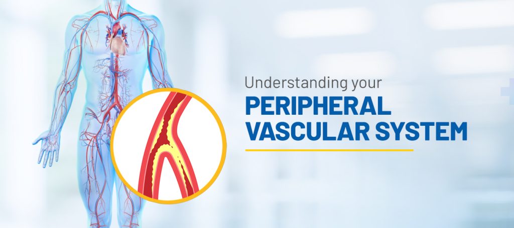
The Peripheral Vascular System is part of the body’s circulatory system which supplies to & receives blood from all parts of the body except Heart and Brain. The Peripheral Vascular system consists Peripheral Arteries which are responsible for oxygenated blood to get supplied from heart to all body parts, and Peripheral Veins carry deoxygenated blood from the extremities back to the heart.

What is peripheral vascular disease?

Peripheral Vascular Disease or PVD is a blood circulation disorder.

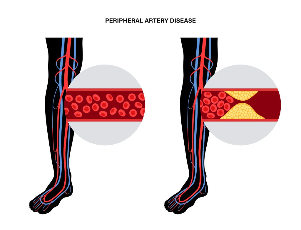
In this condition, your blood vessels narrow or get blocked or spasmed outside the heart and brain.

Peripheral vascular disease can happen in both arteries and veins throughout the body, but often affects the leg’s arteries the most causing fatigue particularly during exercise. This is called intermittent claudication.

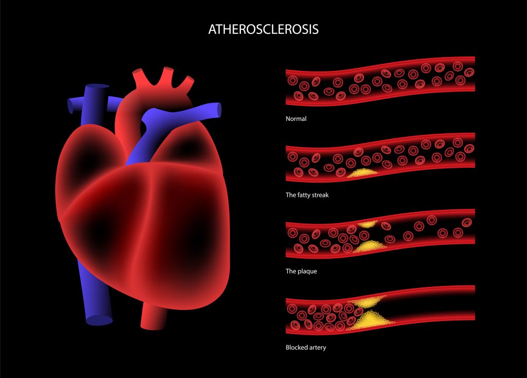
One of the major causes of it is arteriosclerosis which is Plaque building up in a vessel and limiting blood flow and oxygen. As plaque build up progresses, it can reduce or completely block the blood flow in that artery leading to organ damage, loss of fingers, toes, etc.

This condition is different from Coronary Artery Disease and Cerebrovascular disease; these are when the narrowing happens in the heart or the brain respectively.

The Types of PVD

There are two types of Peripheral vascular disease: Functional and Organic

Functional PVD

Vessels widen and narrow in response to brain signals, temperatures changes, etc causing a decrease in the blood flow when vessels are narrowing.

 There is no physical damage to your blood vessels’ structure in functional PVD

The common causes of Functional PVD are emotional distress, colder temperatures, operating vibrating machinery or tools, drugs.

Organic PVD

This is caused due to Physical damage like plaque Built up, inflammation and more. There is damage to the physical blood vessels’ structure. The primary causes of Organic PVD are diabetes, high cholesterol levels, smoking and high blood pressure.

Risk factors

Some lifestyle factors majorly affect your body, hence putting you at a higher risk for PVD. For instance,

  • Overweight,
  • Age above 50,
  • History of stroke or cerebrovascular disease,
  • Abnormal cholesterol levels,
  • Family history of
    • High cholesterol
    • Heart disease,
    • Blood pressure
    • PVD
    • Kidney disease on hemodialysis.

Symptoms of Peripheral vascular disease

For majority, the first sign of PVD is fatigue and discomfort in your legs and feet, which gradually gets worse due to physical activity and lack of blood flow. This sign is rather slow and irregular but persistent.

The major symptom is Claudication. Claudication is lower limb muscle pain experienced while walking. You may notice the pain increasing when you walk long distances or faster and the pain subsides after some rest, but when the pain returns it takes some time to recede.

Other than the pain, some other symptoms also include

  • Reduced hair growth in the legs,
  • Cramps while lying in bed,
  • Arms or legs turning reddish-blue or even page,
  • Legs feeling thin,
  • Paler skin,
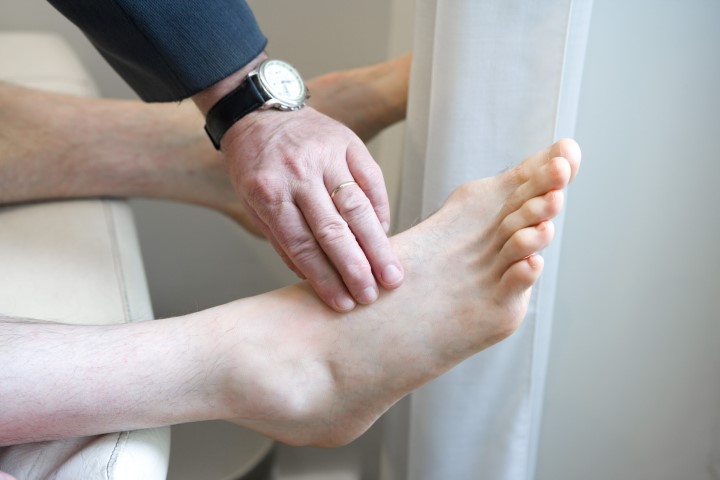
  • Weak pulses,
  • Wounds or ulcers that don’t heal,
  • Thick opaque toenails, and
  • Numbness and heaviness in the muscles.

If you’re experiencing these symptoms, do visit a doctor for a further look. More often than not these symptoms are disregarded as a sign of old age, but delayed diagnosis can lead to complications.

In extreme cases of blood loss, dead tissue can occur. If you suddenly develop a pale, cold limb with weak to no pulse, this is a medical emergency and you require treatment as soon as possible. Untreated limbs as such can lead to amputation.

Treatments for PVD

The treatments for PVD are directed to lower your risk of serious complications and manage your pain and symptoms helping you remain active.

 Major lifestyle modifications  – Initially treatment.

  • Regular balanced diet,
  • Exercise daily
  • Losing some weight.
  • Discontinuing Smoking as it directly causes the reduced blood flow in the vessels, and increases your risk of heart attack or stroke.

Medication

Prescribed by your doctor in order to treat the symptoms.

For instance, medications could be suggested to

  • Increase blood flow and relieve symptoms of leg pain
  • Reduced blood clotting,
  • Lowering high levels of cholesterol and for diabetes may be prescribed

Angioplasty or Vascular surgery

Intervention recommended in cases major artery blockages are diagnosed.

  • Angioplasty is when your doctor inserts a catheter into the artery and a balloon is inflated and open into the artery. In some other cases, a stent is used to keep the artery open.
  •  Vascular surgery, on the other hand, allows the blood to pass the narrow arteries through vein grafting.

If PVD is diagnosed in time, many cases will respond to lifestyle treatments. The best way to examine whether improvement has taken place is to see how far you can walk without pain.

Conclusion

 It is imperative to contact your doctor for a clear diagnosis if your legs look pale or blue to you if your legs become cold or you experience chest pain along with leg pain if you find new sores or ulcers have developed but they don’t heal, fever or any other sign of infections.