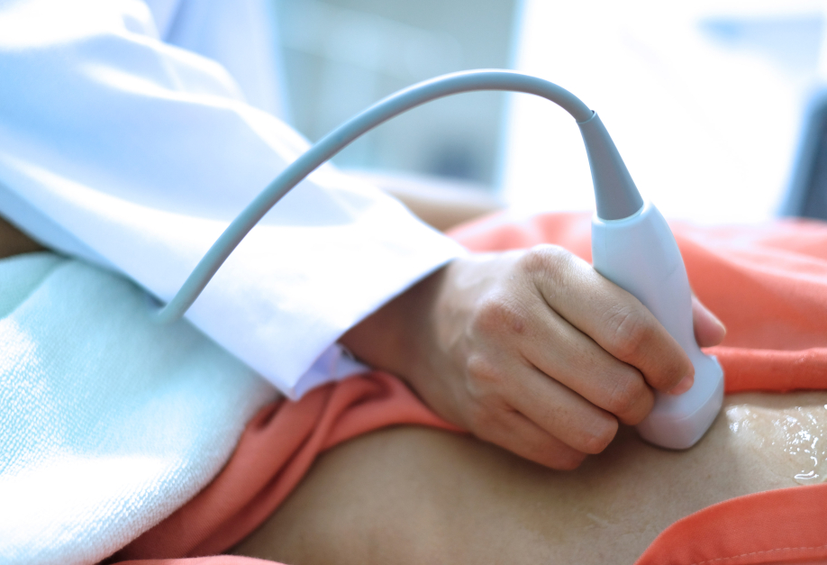
An aorta is the largest artery in the human body supplying clean, oxygenated blood to the entire circulatory system. Aortic valve stenosis is a condition where the aorta of the heart gets blocked. An experienced cardiologist can easily diagnose the disease either through clinical examination, sound wave heart imaging, CT angiography, or other diagnostic modalities.
1) Blood test
2) ECG
3) Echocardiogram
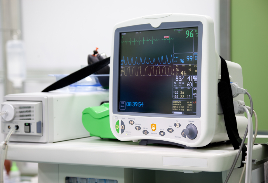
4) Heart Beat Monitoring
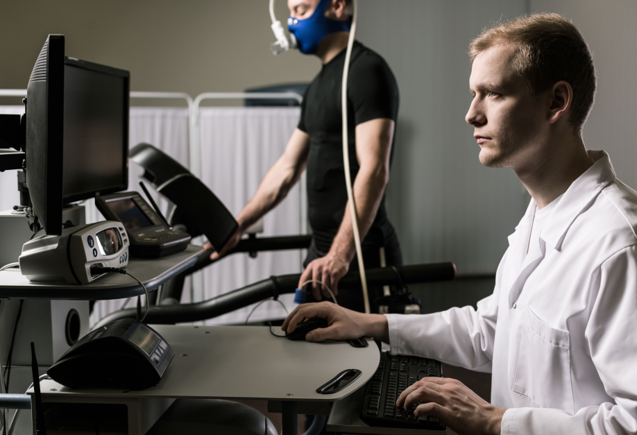
5) Stress Test
Medications can be prescribed to improve the heart’s ability to pump blood which may help compensate for a valve that isn’t working correctly. But, a diseased heart valve being mechanical trouble, cannot be solved by way of medicine alone. Surgery is frequently required to fix the damaged valve. As soon as a conclusion is drawn that the diseased valve needs surgical treatment, valve repair or replacement are the only available options of treatment. To know more, visit your doctor today.
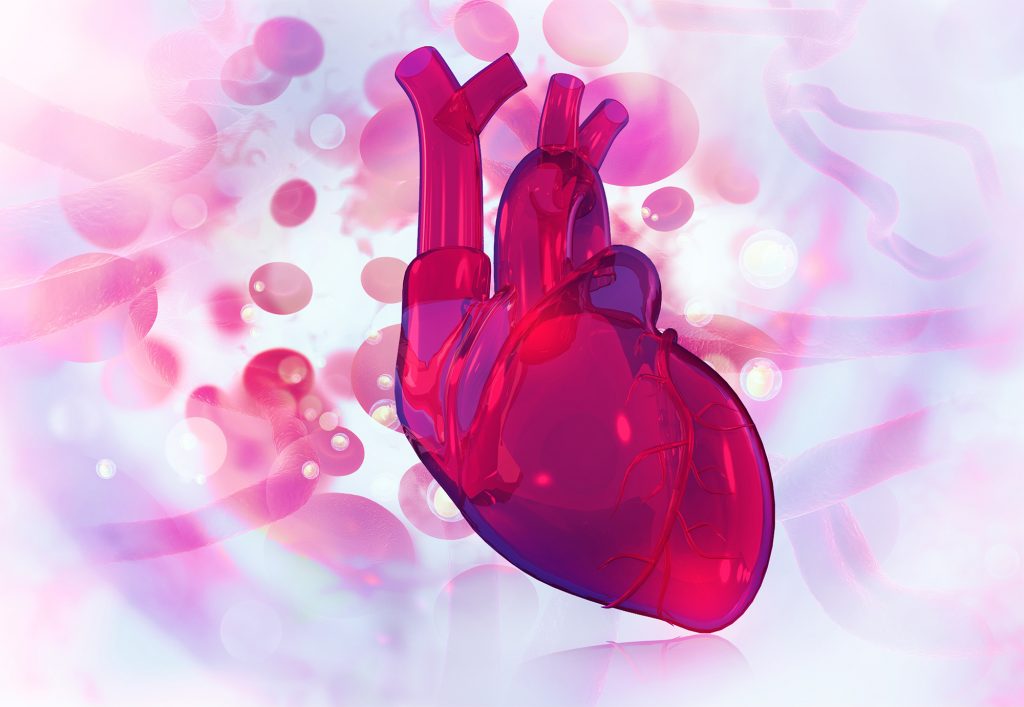
The word aortic stenosis may seem alien, but the condition is one of the most common valvular heart diseases in the world. Usually seen in the elderly, with symptoms surfacing mostly after the age of sixty, Aortic stenosis may also be a congenital condition. Our heart contains four valves, the mitral, tricuspid, pulmonary and the aortic. The former two control the flow of blood from one chamber to another while the pulmonary and aortic direct the blood away from the heart. The pulmonary artery directs the blood to the lungs and the aortic artery directs the blood to the rest of the body. In this condition, an aortic valve begins to show signs of stenosis or narrowing, thus reducing its operating capacity. This results in a reduced supply of oxygenated blood to the body which leads to symptoms like breathlessness, chest pain, fainting and palpitations. If the symptoms are mild or not present, the cardiologist may decide to simply monitor and follow up. However, if the stenosis has shown significant progress and has started to exhibit major symptoms, then the best option is to undergo Transcatheter Aortic Valve Replacement (TAVR).
TAVR is a minimally invasive surgery, as in, the
chest bones are not opened up as in a bypass. It is instead a lot like a stent
placement procedure. Small incisions are made in one’s groin to access the
femoral artery and a hollow tube is placed in it. The new valve is placed on a
tube with a balloon tip on one end and crimped to fit the hollow tube. The
hollow tube is then inserted into the femoral artery and pushed through until
it reaches the diseased valve. At this point, the balloon is inflated which in
turn expands the new valve and helps it take its place. The new valve pushes
the leaflets of the original valve to secure its place. Once the new valve is
up and running, the balloon is deflated and removed.
Transcatheter Aortic Valve Replacement is
relatively new. As a result, it is advised only for high and immediate risk
patients by regulatory authorities and has shown promise in treating patients
at low risk too. As shown by extensive research, heart valve replacement is a
safe and suitable option for elderly patients, with recent studies demonstrating a 94% [1][2] survival rate in 5 years. The survival rate for
any individual patient also depends on other factors like their overall and
cardiac health, co-morbid medical conditions and age. While early TAVR
procedures demonstrated success rates of merely 70-80%, advanced cardiac care,
surgical technologies and tools have increased the rates to as high as 92%. The
overall procedure is also deemed to be nominally risky with a 2% chance of complications
and fatalities across all age groups. This holds true even for elderly patients
above 80.
The recovery process for Transcatheter Aortic
Valve Replacement can take quite a few weeks. Most cardiologists recommend a
course of blood thinners to prevent clots and antibiotics to prevent infection
for their patient’s optimum recovery. Patients can expect to resume their
normal activities gradually once they are totally recovered but will still have
to undergo regular check-ups from their cardiologist. These check-ups are vital
as they are meant to ensure that the patient’s heart and new valve are working
properly.
When it comes to one’s heart, any risk is too big
to take. For those whose lives have been restricted by aortic stenosis,
Transcatheter Aortic Valve Replacement therapy provides an opportunity to lead
a normal life with normal activities again. With experienced surgeons, advanced
tools and techniques, this minimally invasive surgery is sure to change the
lives of those who are suffering from the advanced stages of the disease.
Your heart has four valves that pumps the blood throughout the body. These four valves work relentlessly to ensure blood supply to all the organs in the body. But sometimes as time passes, the usual wear and tear sets in causing the main valve, called the aortic valve in the heart to be blocked.
Once blocked it becomes very difficult for the heart to pump the adequate amount of blood required by the body resulting in breathlessness, chest-pains and finally heart attacks.
The patient needs to undergo valve replacement surgery to solve/treat this disease. Usually, valve replacements are done through open-heart surgery wherein a new tissue or mechanical prosthetic valve is inserted in the place of the diseased valve.
Open heart surgery is an invasive procedure with a very long recovery period. This can be a difficult challenge for patients, especially elderly patients.
But there is an alternate method to open heart surgery which is less invasive and has a shorter recovery period. It’s called a TAVI.
This minimally aggressive procedure includes positioning a novel artificial valve over the diseased native valve of the patient through a catheter introduced through the patient’s large artery located in the groin (femoral artery).
This procedure can thus be done through small openings and hence result in quicker recovery post procedure enabling the patients to go back to their normal lives.
To know more about the procedure, visit your cardiologist today.